Next-Generation Biomarkers in Bladder Cancer: Urinary and Tissue Signatures
Abstract
Bladder cancer is among the most common malignancies of the urinary tract and remains a major contributor to global cancer-related morbidity and mortality. Despite advances in diagnosis and treatment, its high recurrence rate necessitates lifelong surveillance. Although cystoscopy and urine cytology remain the diagnostic cornerstone, their inherent limitations have driven the development of urinary and tissue biomarkers to improve early detection, surveillance, prognostic assessment, and prediction of treatment response.
Several FDA- approved urinary biomarkers-including Nuclear Matrix Protein 22 (NMP22), Bladder Tumor Antigen (BTA), UroVysion Fluorescence in Situ Hybridization (FISH), and ImmunoCyt/uCyt+ immunofluorescence have demonstrated varying diagnostic performance. Simultaneously, advances in genomics, transcriptomics, proteomics, metabolomics, epigenetics, extracellular vesicles, and liquid biopsy technologies have considerably expanded the landscape of bladder cancer diagnostics. Furthermore, artificial intelligence-assisted biomarker integration represents an emerging frontier that may enhance precision diagnostics and individualized patient care.
This narrative review comprehensively summarizes established and emerging urinary and tissue biomarkers in bladder cancer, critically evaluates their clinical utility and limitations, and discusses future directions toward precision medicine and personalized management in urothelial carcinoma.
Keywords
Bladder cancer; Urothelial carcinoma; Biomarkers; Urine cytology; Carcinoma in situ; Liquid biopsy; Precision medicine; Urinary biomarkers
Abbreviations
BC: Bladder Cancer; NMIBC: Non-Muscle-Invasive Bladder Cancer; MIBC: Muscle-Invasive Bladder Cancer; CIS: Carcinoma in situ; FISH: Fluorescence in situ Hybridization; NMP22: Nuclear Matrix Protein 22; BTA: Bladder Tumor Antigen; ctDNA: Circulating Tumor DNA
Introduction
Bladder cancer is one of the most frequently diagnosed malignancies of the genitourinary tract and remains a major global healthcare burden [1,2]. According to recent epidemiology estimates, bladder cancer ranks among the ten most common cancers worldwide, accounting for significant morbidity and mortality, particularly among older adults and males [2]. Urothelial carcinoma represents nearly 90% of bladder malignancies in developed countries and demonstrates marked biological heterogeneity, ranging from indolent Non-Muscle-Invasive Bladder Cancer (NMIBC) to highly aggressive Muscle-Invasive Bladder Cancer (MIBC), thereby contributing to substantial variability in prognosis and therapeutic outcomes [1,2].
One of the most clinically challenging characteristics of bladder cancer is its remarkably high recurrence rate, necessitating repeated surveillance and prolonged follow-up in many patients [1,3]. Approximately 50% to 70% of patients with NMIBC experience disease recurrence following initial treatment, while a subset eventually progresses to muscle-invasive disease despite intervention [4,5]. Consequently, bladder cancer is recognized as one of the most expensive malignancies to manage on a per-patient basis from diagnosis to death because of repeated cystoscopic evaluations, urinary testing, imaging studies, and therapeutic interventions [1,6].
Currently, cystoscopy remains the gold standard for diagnosis and surveillance of bladder cancer [4]. Despite its established diagnostic role, cystoscopy is invasive, costly, associated with patient discomfort, and may negatively affect compliance during long-term surveillance programs [4]. Additionally, certain lesions-particularly flat lesions such as Carcinoma in Situ (CIS)-may occasionally remain difficult to detect using conventional cystoscopic assessment alone, emphasizing the need for supplementary diagnostic strategies [4].
Despite its longstanding role in bladder cancer evaluation, urine cytology remains constrained by variable diagnostic performance, particularly in the detection of low-grade urothelial tumors [4,5]. While the test offers excellent specificity and maintains important clinical value in identifying high-grade malignancies and Carcinoma in Situ (CIS), its limited sensitivity reduces reliability for early-stage and low-grade disease [4,5]. Consequently, reliance on urine cytology alone may result in missed diagnosis in selected patient populations, reinforcing the need for more sensitive, noninvasive diagnostic approaches capable of improving detection and surveillance [5,6].
Recent advances in molecular biology and precision oncology have substantially expanded bladder cancer biomarker research [6-8]. FDA-approved urinary biomarkers-including Nuclear Matrix Protein 22 (NMP22), Bladder Tumor Antigen (BTA), UroVysion Fluorescence in Situ Hybridization (FISH), and ImmunoCyt/uCyt+ immunofluorescence have demonstrated variable diagnostic utility and are currently used as adjunctive tools in selected clinical scenarios [5,6]. Moreover, novel genomic, transcriptomic, epigenetic, proteomic, metabolomic, extracellular vesicle, and liquid biopsy approaches have emerged as promising strategies for improving diagnostic accuracy, recurrence prediction, prognostication, and treatment personalization [6,8].
Given the limitations of conventional diagnostic modalities and the expanding landscape of molecular diagnostics, a comprehensive evaluation of bladder cancer biomarkers is warranted. In this narrative review, we discuss established and emerging urinary and tissue biomarkers in bladder cancer, critically evaluate their diagnostic and prognostic utility, examine current limitations, and explore future directions towards precision medicine and individualized management in urothelial carcinoma.
Current Diagnostic Approaches and Limitations in Bladder Cancer
Bladder cancer diagnosis and surveillance continue to rely primarily on cystoscopy, urine cytology and radiological imaging. Although these modalities remain central to current clinical practice, each possesses important limitations that have stimulated the development of urinary and tissue biomarkers. Conventional white-light cystoscopy is regarded as the gold standard for bladder cancer detection because it allows direct visualization of the bladder mucosa and facilitates histopathological confirmation through biopsy or transurethral resection. However, cystoscopy is invasive, costly, uncomfortable for patients, and may fail to identify flat lesions such as Carcinoma in Situ (CIS). In addition, the need for repeated cystoscopic examinations during long-term surveillance significantly increases healthcare costs and negatively affects patients’ quality of life [1,7-9].
Urine cytology is commonly used as an adjunctive diagnostic tool because of its excellent specificity for high-grade urothelial carcinoma and CIS. Nevertheless, its sensitivity for low-grade tumors remains poor, limiting its role as a reliable standalone screening or surveillance test. Furthermore, cytological interpretation may vary according to specimen quality and observer expertise [6,7,10].
Imaging modalities including Computed Tomography Urography (CTU) and multiparametric Magnetic Resonance Imaging (mpMRI) provide valuable information regarding upper urinary tract involvement, local staging, and muscle invasion. The development of the Vesical Imaging-Reporting and Dat System (VI-RADS) has improved MRI standardization in bladder cancer staging. Despite these advances, imaging techniques have limited sensitivity for small papillary lesions and microscopic recurrence, making them unsuitable replacements for cystoscopy in routine surveillance [11,12].
To overcome the limitations of conventional diagnostic methods, several enhanced cystoscopic technologies such as Narrow-Band Imaging (NBI) and Photodynamic Diagnosis (PDD) have been introduced. These approaches improve visualization of flat and small lesions and may reduce recurrence following transurethral resection. However, their widespread implementation remains limited by cost, equipment availability, and procedural complexity [13].
Taken together, current diagnostic pathways leave an important clinical gap in bladder cancer management. Although cystoscopy remains indispensable, its invasiveness and limitations have created growing interest in non-invasive biomarkers capable of improving diagnosis, surveillance, risk stratification, and prediction of treatment response. Consequently, urinary biomarkers have emerged as promising adjunctive tools in modern Uro-oncology practice.
Urinary Biomarkers
FDA-approved and commercially available urinary biomarkersSeveral urinary biomarkers have been developed to improve the detection and surveillance of bladder cancer. Some assays have received regulatory approval and are currently used as adjuncts to cystoscopy and urine cytology in selected clinical settings.
NMP22Nuclear Matrix Protein 22 (NMP22) is one of the most extensively studied urinary biomarkers in bladder cancer. NMP22 is released from apoptotic and necrotic urothelial tumor cells and can be detected in urine using quantitative ELISA or point-of-care assays BladderChek format. Across studies, diagnostic performance is variable and case-mix dependent; a meta-analysis of BladderChek reported pooled sensitivity of 56% and specificity 88%, while broader reviews generally place NMP22 in a reported range of roughly 55% to 70% sensitivity and 70% to 88% specificity. The assay’s main advantages are operational simplicity, rapid turnaround, and sensitivity that may exceed cytology in some lower-stage settings. Its major weakness is, specificity: urinary tract infection, urolithiasis, inflammation, hematuria, other benign urothelial injury or recent instrumentation can all produce false-positive results [6,14,15]. Despite these limitations, NMP22 may serve as a useful adjunctive test in selected patients undergoing bladder cancer surveillance.
BTA Stat and BTA TRAKThe BTA assay’s detect human complement factor H-related protein in urine. BTA stat is a qualitative office-based immunochromatographic test, whereas BTA TRAK is quantitative and ELISA-based. Their attraction is practical convenience, but performance is highly vulnerable to biological confounding. Review-level data suggest that BTA stat has a reported sensitivity around 57% to 83% and specificity around 60% to 92%, while BTA TRAK has reported sensitivity around 66% to 77% and specificity around 45% to 81%. Inflammatory conditions, gross or microscopic hematuria, stones, recent BCG, and other benign urinary tract insults can markedly reduce specificity. Consequently, BTA assay’s may outperform cytology in raw sensitivity, but the specificity penalty often limits their usefulness for confident rule-in or rule-out decision-making. In contemporary practice, they are more important as historical comparators than as universally preferred assays [6,16,17].
UroVysion FISHUroVysion FISH detects chromosomal abnormalities that are common in urothelial carcinoma, classically involving chromosomes 3,7, and 17 together with 9p21 deletion. Compared with protein tests, its appeal is greater disease specificity at a still reasonable sensitivity. Pooled reviews generally place UroVysion sensitivity in the low-to-mid 70% range and specificity around the low-to-mid 80% range. Clinically, UroVysion is most useful when cytology is atypical or equivocal, when CIS or high-grade disease is suspected, or when clinicians want a more molecular adjunct to conventional surveillance in higher-risk patients. Its disadvantages are cost, laboratory dependency, technical complexity, and the interpretive challenge of discordant results, especially positive FISH with negative cystoscopy. Such discordance may precede visible recurrence, but it can also generate additional testing without immediate actionable disease [6,16,17].
ImmunoCyt and uCyt+ImmunoCyt/uCt+ is an immunofluorescence assay that targets exfoliated urothelial cells using antibodies directed against CEA and mucin-like antigens. Across pooled analyses, sensitivity is usually higher than cytology, commonly in the range of 73% to 84%, whereas specificity is lower, often around 62% to 75%. This gives the test a recognizable profile: it is more useful as a sensitivity-enhancing adjunct than as a stand-alone discriminator. In practice, ImmunoCyt can be helpful when cytology is suspicious but not definitive, or when clinicians are particularly concerned about missing high-grade recurrence. The limitations are also familiar: lower specificity, dependence on specialized processing, reduced availability, and less consistent integration into modern pathway than might be expected from its historical performance profile [6,16,17].
CxbladderCxbladder is a commercially available multigene urinary mRNA assay that uses multigene urinary mRNA signatures, with different assay versions designed for detection, triage, or surveillance. In the original hematuria-development study, the finalized Cxbladder algorithm achieved 82% sensitivity at 85% specificity, while detecting 97% of high-grade tumors and all tumors stage T1 or greater. Most recently, the STRATA microhematuria trial reported that Cxbladder Triage yielded 90% sensitivity, 56% specificity and 99% negative predictive value for urothelial carcinoma. The principal clinical value of the platform is therefore not high specificity, but strong negative prediction in selected populations; this makes it attractive when the aim is to reduce unnecessary cystoscopy in appropriately counseled patients. The caveats are that performance varies by assay version and population, independent head-to-head validation remains less extensive than for legacy tests, and regulatory status differs from the FDA-cleared marker group. For a balanced narrative review, Cxbladder is best presented as a clinically relevant commercial molecular assay with promising workflow utility, especially in hematuria triage rather than as a universal cystoscopy substitute [16-18].
Table 1 summarizes the practical distinctions among the best-known urinary assays. Because diagnostic estimates depend strongly on setting, the values shown are representative reported ranges or landmark study values rather than universal constants [16-20].
Cxbladder is included because of clinical relevance and current workflow use, although manufacturer labelling states that it is a CLIA laboratory-developed test rather than an FDA-cleared assay [18].
Table 1: Comparison of established urinary biomarkers in bladder cancer.
| Assay & Method | Target | Representative Performance | Most Defensible Clinical Role | Main Advantages | Main Limits |
|---|---|---|---|---|---|
| NMP22 | Nuclear Matrix Protein 22; ELISA or point-of-care immunoassay |
Sensitivity: ~55%–70%; Specificity: ~70%–88% |
Adjunctive triage or supportive surveillance test in some settings | Rapid, simple; higher sensitivity than cytology | False positives with infection, stones, etc. |
| BTA stat / BTA TRAK | Human complement Factor H-related protein; qualitative or quantitative immunoassay |
BTA stat sensitivity: ~57%–83%, specificity: ~60%–92%; BTA TRAK sensitivity: ~66%–77%, specificity: ~45%–81% |
Limited adjunctive use; historical comparator | Office convenience, easy workflow | Markedly affected by hematuria, BCG, etc. |
| UroVysion FISH | Aneuploidy of chromosomes 3/7/17 and 9p21 loss in exfoliated cells |
Sensitivity: ~72%–76%; Specificity: ~83%–85% |
Adjudication of equivocal cytology; higher-risk surveillance; suspected CIS; difficult cases | Better molecular specificity than protein markers | Expensive, labor intensive; not ideal for routine universal use |
| ImmunoCyt/uCyt+ | Immunofluorescent detection of CEA and mucin-like antigens on exfoliated cells |
Sensitivity: ~73%–84%; Specificity: ~62%–75% |
Sensitivity-enhancing adjunct, especially with equivocal cytology | Good sensitivity, especially for higher-grade disease | Lower specificity; specialized processing; limited contemporary uptake |
| Cxbladder | Urinary multigene mRNA RT-qPCR panels |
Original study: 82% sensitivity, 85% specificity; STRATA Triage: 90% sensitivity, 56% specificity, 99% NPV |
Hematuria triage; selected surveillance de-intensification workflows | High NPV; clinically attractive rule-out profile | Different assay versions; performance depends on indication; regulatory category differs from FDA-cleared legacy tests |
Cxbladder is included because of clinical relevance and current workflow use, although manufacturer labelling states that it is a CLIA laboratory-developed test rather than an FDA-cleared assay [18].
Emerging Urinary Biomarkers in Bladder Cancer
Among emerging urinary biomarkers, the most mature signals currently come from DNA methylation and urinary tumor-DNA/cell-free DNA because they exploit stable disease-proximal molecular alterations and increasingly show the high negative predictive value needed for surveillance de-intensification. By contrast, miRNA, exosome, proteomic, and metabolomic approaches remain biologically compelling but more heterogeneous, and their immediate value is most likely in triage, surveillance support, and risk enrichment, rather than outright replacement of cystoscopy [19,20].
DNA methylation biomarkersUrinary DNA methylation assays are attractive because hypermethylated CpG patterns are chemically stable, detectable in exfoliated urothelial DNA, and often arise early in tumorigenesis. Representative platforms include Bladder EpiCheck and newer PCR-based targeted methylation panels developed for hematuria triage and pre-operative risk stratification. In NMIBC surveillance, Bladder EpiCheck reported 68.2% sensitivity, 88.0% specificity, and 95.1% NPV, with 99.3% NPV when low-grade Ta recurrences were excluded; in hematuria/suspected-cancer cohorts, other methylation panels have reported sensitivities and specificities in the mid- to high- 80% range, sometimes exceeding 90% sensitivity in validation cohorts [21]. Clinically, methylation is best positioned for rule-out triage, surveillance support, and possibly risk stratification before definitive endoscopy. Its main limitations are assay-to-assay heterogeneity, variable thresholds, incomplete head-to-head validation against established commercial tests, and uncertainty about performance in benign inflammatory states and diverse populations. The key open question is whether a locked methylation panel can safely defer cystoscopy in selected low-risk pathways without missing clinically meaningful high-grade disease.
MicroRNA biomarkersUrinary miRNAs are biologically plausible because they reflect post-transcriptional dysregulation in bladder carcinogenesis and remain relatively stable in urine, especially when packaged in vesicle or measured from cell pellets. Most assays rely on qRT-PCR of single miRNAs or multimarkers panels. A recent meta-analysis found that urinary miRNAs achieved an AUC of 0.88, with 82% sensitivity and 81% specificity; for the recurrently studied miR-143, pooled performance was 79% sensitivity and 87% specificity, while multimarker panels generally outperformed single-analyte assays [22]. This profile makes miRNAs more attractive for initial diagnosis/triage than for stand-alone surveillance. They may also contribute prognostic information, since some panels correlate with recurrence, progression, and treatment resistance. However, translation is slowed by inconsistent preanalytics, different analyte sources (whole urine, sediment, cell-free fraction, exosome-enriched fraction), and unresolved normalization strategies. The main open questions are which endogenous controls are reproducible across laboratories and whether urinary miRNA adds incremental value once methylation and ctDNA are already available in the same workflow.
Extracellular vesicles and exosomesExtracellular vesicles, especially exosomes, are appealing because bladder tumors release them directly into urine, and their lipid membrane protects RNA and protein cargo from degradation. Current assay’s focus mainly on exosomal lncRNAs, miRNAs, and mixed ncRNA panels. In a 2024 urine-specific meta-analysis, urine-derived exosomes showed pooled 75% sensitivity, 77% specificity, and AUC 0.83 for bladder cancer diagnosis; multidimensional exosome panels outperformed single-cargo tests, with subgroup AUCs approaching 0.87 [23]. Representative lncRNA-based models have also shown validation-set AUCs around 0.85. Thus, the most plausible role of exosomes is noninvasive diagnosis and possibly prognostic enrichment, especially if multimarker cargo panels are used. The main barriers are technical rather than conceptual: isolation methods are not standardized, yield and purity vary by kit and centrifugation workflow, and turnaround remains less practical than PCR-based methylation or targeted DNA assays. The critical open question is whether simplified, kit-based exosome workflows can preserve signal quality in real-world multicenter practice.
Urinary ctDNA and cfDNAUrinary circulating tumor DNA (ctDNA) and cell-free DNA (cfDNA) arguably has the strongest mechanistic link to bladder cancer because urine is the fluid most proximal to the tumor, carrying tumor-derived mutations, copy-number changes, methylation abnormalities, and fragmentation patterns. Representative assays include targeted deep sequencing platform such as uCAPP-Seq, hotspot mutation panels for genes such as TERT and FGFR3, and newer cfDNA fragmentomics models. In the landmark uCAPP-Seq study, pretreatment urine tumor DNA detection reached 93% sensitivity with tumor-informed profiling and 84% sensitivity in a tumor-naïve approach, both with 96% to 100% specificity; during surveillance, utDNA identified 91% of patients who later recurred and preceded clinical progression in 92% of these cases [24]. Multiomic urine cfDNA analysis before cystectomy also detected molecular residual disease with about 81% sensitivity and 81% specificity for non-pCR and predicted survival [25]. More recently, a cfDNA fragmentation-hotspot machine-learning model achieved AUC 0.96 and 87% sensitivity at 100% specificity, including 71% sensitivity in early-stage disease. This class therefore has credible use in diagnosis, surveillance, Minimal Residual Disease (MRD) detection, prognosis, and potentially treatment selection. The main limitations are cost, sequencing infrastructure, low tumor fraction in some low-grade lesions, and the need to distinguish actionable recurrence from urothelial field effects.
Proteomic biomarkersProteomic biomarkers reflect a different layer of tumor biology: altered cell-cycle activity, extracellular-matrix remodeling, inflammation, and aberrant glycosylation. The most clinically recognizable representative is ADXBLADDER, a urinary MCM5- based assay that measures MCM5 in urinary sediment, alongside broader exploratory protein/glycoprotein candidates such as BLCA-4, HTRA1, keratin 17, MMP9, and glycoform-based signatures. In hematuria evaluation, ADXBLADDER outperformed cytology in a large multicenter study; across studies summarized in recent reviews, proteomic tests in this space generally show 45% to 73% sensitivity and 74% to 100% NPV, with stronger performance for higher-grade disease [26,27]. This suggests that the best current role for proteomics is adjunctive triage or surveillance rule-out, not definitive diagnosis. The main limitation is biological noise: urinary proteins are heavily influenced by inflammation, hematuria, proteinuria, instrumentation, and assay batch effects. The important open question is whether future glycoproteomic assays can outperform simple protein-concentration tests by capturing cancer-specific post-translational changes rather than nonspecific tissue injury.
Metabolomic biomarkersMetabolomic is conceptually attractive because bladder cancer rewires glycolysis, amino-acid use, nucleotide turnover, and lipid metabolism, and these changes may be directly measurable in urine. Representative platforms include UPLC-MS, LC-MS/MS, and NMR-based signatures containing 5 to 11 metabolites. Reported performance remains wide: an older urinary metabolomic model for diagnosis and survival prediction showed approximately 78% sensitivity and 70.3% specificity in validation, whereas a more recent 11-metabolite UPLC-MS panel reported 95.3% sensitivity and 100% specificity in its study framework [28]. At present, the best use case is probably diagnostic enrichment in hematuria or combination with other molecular assays rather than stand-alone decision-making. The major limitations are strong sensitivity to diet, smoking, medication exposure, renal function, and sample handling, together with incomplete harmonization of metabolite identification pipelines. The key open question is whether metabolomic signatures retain accuracy in external multicenter cohorts enriched for common confounders seen in routine urology clinics.
AI and multi-omics integrationArtificial Intelligence (AI) is best understood not as a separate biomarker class, but as the layer that selects features, fuses data types, and converts multi-signal urine testing into a clinically usable probability of meaningful disease. The strongest examples already come from urinary DNA analysis: the cfDNA approaches that combine targeted sequencing with low-pass whole-genome information have shown value for MRD detection and survival prediction [29]. Reviews of AI in bladder cancer emphasize that these tools are promising for detection, staging, prognostication, and therapy selection, but remain mostly retrospective and insufficiently calibrated for routine deployment [30]. The most realistic near-term application is layered clinical decision support: molecular triage before first cystoscopy, high-NPV surveillance between cystoscopies, and molecular escalation when recurrence risk or treatment resistance is suspected. The open questions are model transportability, calibration drift, interpretability, and how best to combine biomarkers with age, smoking, hematuria type, imaging, and prior pathology.
The Table 2 below synthesizes representative ranges and intended roles from the studies cited above.
Table 2: Urinary biomarkers in bladder cancer.
| Biomarker Class | Representative Assays | Typical Performance Ranges | Best Clinical Role | Main Limitations |
|---|---|---|---|---|
| DNA Methylation | Bladder EpiCheck targeted PCR methylation panel |
Sensitivity: ~68–91%; Specificity: ~86–90%; NPV often >95% in surveillance |
Triage, surveillance support, risk stratification | Panel heterogeneity, thresholding, need for prospective studies |
| miRNA | qRT-PCR single miRNAs; multi-miRNA panels |
Sensitivity: ~79–82%; Specificity: ~81–87% |
Diagnosis/triage; adjunct prognosis | Normalization, specimen-source variability, poor standardization |
| Extracellular Vesicles/Exosomes | Exosomal lncRNA/miRNA panels |
Sensitivity: ~75–78%; Specificity: ~77–81%; AUC ~0.83–0.87 |
Diagnosis; biomarker discovery for prognosis | Isolation variability, labor intensity, contamination, platform complexity |
| Urinary ctDNA/cfDNA | uCAPP-Seq; hotspot mutation panels; fragmentation models |
Sensitivity: ~84–93% in diagnosis; Specificity: ~96–100%; MRD sensitivity/specificity ~81/81 |
Diagnosis, surveillance, MRD, prognosis, treatment guidance | Cost, sequencing depth, field-effect biology, reimbursement |
| Proteomic Biomarkers/Glycoprotein Panels | ADXBLADDER/MCM5; protein/glycoprotein panels |
Sensitivity: ~45–73%; NPV: ~74–100% |
Adjunctive triage and surveillance rule-out | Biological noise from inflammation/hematuria, limited assay standardization |
| Metabolomic Biomarkers | UPLC-MS, LC-MS/MS, NMR metabolite panels |
AUC: ~0.69–0.98; Sensitivity/specificity vary widely |
Triage; combination panels | Strong confounding from diet, smoking, renal function, sample handling |
| AI and Multi-Omics Integration Models | ML-based fragmentomics; multi-omic cfDNA integration studies | AUC up to ~0.96 in discovery/validation studies | Decision support across triage, surveillance, treatment selection | External validation, calibration, explainability, workflow integration |
Tissue Biomarkers in Bladder Cancer
Several tissue biomarkers inform bladder cancer biology and management. FGFR3 mutations occur in ~60% of low-grade NMIBC and ~15-20% of MIBC [31,32], defining a luminal-papillary subtype with relatively favorable prognosis. These alternations are targetable: the FGFR inhibitor erdafitinib produced ~40% response rates in FGFR3-altered advanced disease and earned FDA approval [33]. TP53 mutations are frequent (~60-90%) in high-grade/basal tumors and neuroendocrine variants [33], marking aggressive behavior and poor outcome. A high ki-67 proliferation index similarly correlates with recurrence risk, especially in T1 tumors. PD-L1 expression (by IHC) is a predictive biomarker for immune checkpoint therapy, guiding first-line use of pembrolizumab or atezolizumab in cisplatin-ineligible metastatic patients [33], though many PD-L1- negative tumors still respond [33]. HER2 (ERBB2) overexpression/amplification occurs in a minority of UC (up to ~10%) [32,33] and may identify candidates for anti-HERS2 agents or ADCs, but single-agent trials have been disappointing. Consensus molecular subtype (basal/squamous, luminal-papillary, etc.) further stratify tumors by biology and inform prognosis [33]. In practice, tissue markers can complement urinary tests by refining risk stratification: e.g. an FGFR3- mutant NMIBC may be observed, whereas TP53-mutant MIBC may prompt early aggressive therapy. For patients with hematuria or surveillance findings, positive molecular results (urine or tissue) would trigger cystoscopy, whereas negative high-precision tests could safely extend follow-up intervals. Despite promise, most biomarkers remain investigational: assays lack standardization, result vary by cohort, and multi-marker models need large prospective validation [33].
FGFR3Activating FGFR3 mutations are characteristics of low-grade NMIBC and luminal-papillary MIBC. In large series, ~60-70% of Ta tumors and ~15-20% of invasive tumors harbor FGFR3 alterations. FGFR3-mutant tumors often exhibit papillary histology and intact p53, and they generally portend a lower progression risk. Importantly, FGFR3 serves as a drug target: the pan-FGFR inhibitor erdafitinib yielded a 40% complete/partial response rate in FGFR3-mutant advanced UC patients previously treated with chemotherapy, leading to FDA approval in 2019. FGFR3 mutation therefore predicts both relatively favorable NMIBC behavior and sensitivity to FGFR blockade (e.g. erdafitinib) [33].
TP53By contrast, TP53 mutations (or p53 overexpression by IHC) are hallmarks of high-grade disease. TP53 alterations are seen in ~50-70% of MIBCs and are enriched in basal/squamous and neuroendocrine-like subtype. In consensus subtype analysis, ~61% of basal/squamous tumors and ~94% of neuroendocrine-like tumors carried TP53 mutations. TP53-mutant UCs show high genomic instability and tend to be more aggressive. While no TP53-targeted drugs are currently approved, TP53 status informs prognosis: mutated TP53 (or high nuclear p53 staining) correlated with higher stage/grade and poorer survival [33].
Ki-67The ki-67 nuclear antigen marks proliferative fraction. Ki-67 labelling indices >20-30% are common in high-grade bladder tumors, and higher Ki-67 by IHC predicts recurrence and progression. Meta-analyses and consensus panels have identified ki-67 as one of the stronger proliferation markers for NMIBC prognosis. For example, tumors with high ki-67 (and concomitant p53 overexpression) have significantly worse recurrence-free survival. Thus, ki-67 helps refine risk beyond grade alone, although exact cutoffs and scoring vary by study.
PD-L1 (CD274)PD-L1 expression by tumor cells or immune infiltrates is a key predictive biomarker for immune checkpoint therapy. Several anti-PD-1/PD-L1 agents (pembrolizumab, atezolizumab, nivolumab, avelumab, durvalumab) are approved for UC. In first-line cisplatin-ineligible metastatic UC, pembrolizumab or atezolizumab monotherapy is indicated only if the tumors PD-L1 score meets a threshold (e.g. Combined Positive score >10 for pembrolizumab). In second line (post-platinum) setting, PD-L1 testing is not required (pembrolizumab showed OS benefit regardless of PD-L1). Critically, the predictive value of PD-L1 is imperfect: many PD-L1 negative patients still respond to ICB, and conversely some PD-L1 positive tumors fail to benefit [33]. Different assays (22C3, SP142, SP263) and scoring (TPS, CPS, IC) further complicate interpretation. In summary, PD-L1 IHC is routinely performed to guide first line immunotherapy in metastatic IC (per FDA/EMA labelling), but its role in NMIBC or surveillance is limited.
HER2 (ERBB2)ERBB2 gene amplification or protein overexpression occurs in a minority of bladder cancers. Estimates vary, about 9% to 12% of invasive UCs show HER2 positivity (higher rates in micropapillary or T4 tumors). HER2 upregulation is seen mostly in luminal-papillary contexts. While early trials of trastuzumab or lapatinib plus chemotherapy showed only modest benefit, novel approaches (HER2- targeted ADCs like T-DM1, trastuzumab deruxtecan) are under study. Notably, one series reported essentially zero HER2+ cases among upper-tract UCs, and overall HER2 alterations appear rare and concentrated in aggressive cases [34]. Thus, routine HER2 testing in UCs is not yet standard, but may identify a small subset for clinical trials.
Molecular subtypesGene-expression profiling has revealed intrinsic subtypes of bladder cancer analogous to breast cancer (luminal vs. basal) plus a neuroendocrine-like class. Luminal subtypes (including luminal-papillary and luminal-infiltrated) express urothelial differentiation markers (GATA3, FOXA1, KRT20) and often carry FGFR3 mutations. Basal subtypes express basal keratins (KRT5/6, KRT14) and have frequent TP53/RB1 alterations [33]. These subtypes have prognostic significance: basal tumors tend to be more aggressive but may be more chemo sensitive, whereas luminal-papillary tumors often recur but progress more slowly. Importantly, consensus classification initiatives have harmonized multiple schema into six classes (LumP, LumNS, LumU, Stroma-rich, Ba/sq, NE-like) with distinct outcomes. Subtype assignment (via panels or RNA-seq) is still largely research–based, but may in future guide therapy selection (e.g. immunotherapy appears most effective in certain luminal subtypes, while Ba/Sq tumors have high immune infiltration) (Table 3).
Table 3: Summary of key tissue biomarkers in bladder cancer.
| Biomarker | Frequency/Prevalence | Clinical Implication | Evidence Level |
|---|---|---|---|
| FGFR3 | ~60–70% of Ta/T1; ~15–20% of MIBC | Luminal-papillary tumors; favorable prognosis; targetable (erdafitinib) | Phase 2/3 trials (FDA-approved use) |
| TP53 (p53) | ~50–70% of MIBC (basal/NE subtypes) | Basal/NE subtypes; aggressive/high-grade; poor prognosis | Retrospective studies; prognostic |
| Ki-67 | Highly expressed in high-grade tumors | High proliferation index predicts recurrence/progression | Numerous cohorts, meta-analyses |
| PD-L1 (IHC) | ~20–30% of advanced UC (varies by assay) | Predictive for checkpoint immunotherapy (required for 1st-line in cisplatin-ineligible); imperfect (not prognostic) | Phase 2/3 trials; regulatory label |
| HER2 (ERBB2) | ~9–12% (mostly high-grade/micropapillary) | Potential target (trastuzumab/ADC); prognostic significance unclear | Early-phase studies; no approved use |
| Subtypes | Consensus classes (LumP, LumNS, LumU, stroma-rich, Ba/Sq, NE) | Reflect biology: LumP (FGFR3+, papillary), Ba/Sq (TP53+, aggressive); inform trial design | Consortia (TCGA, Kamoun et al.) |
Although established tissue biomarkers such as FGFR3, TP53, KI-67, PD-L1, and HER2 continue to play important roles in prognostication and therapeutic decision-making, the future of precision bladder cancer management will likely depend on their integration with next-generation urinary biomarkers, including DNA methylation assays, circulating tumor DNA (ctDNA), extracellular vesicles, and AI-driven multi-omics platforms. This integrated approach has the potential to improve early detection, risk stratification, surveillance, and personalized treatment selection.
Clinical applications of biomarkers- Hematuria evaluation: Noninvasive urinary panels (DNA methylation, mutations, exosomal RNAs) may eventually triage patients with microscopic or gross hematuria. In current practice, a positive high-sensitivity molecular test would prompt expedited cystoscopy, while a robustly negative test (especially in low-risk patients) might safely defer invasive evaluation. For example, a tumor-specific FGFR3 or TERT mutation detected in voided urine could identify an occult cancer [33], whereas absences of any mutation or methylation markers could support surveillance over immediate cystoscopy.
- NMIBC surveillance: Tissue and urinary biomarkers together can stratify recurrence risk. Tissue factors (high-grade, multifocality, presence of carcinoma in situ, TP53 mutation, high ki-67) inform baseline risk and surveillance intervals. Emerging urine tests (e.g. FGFR3 mutations assay, methylation panels, UroSEEK) offer sensitive recurrence detection [33]. In BCG-treated NMIBC, persistent or rising urinary tumor DNA may signal early recurrence before visible lesions. Patients with negative urine markers at follow-up might avoid some routine cystoscopies.
- Recurrence and progression prediction: Tissue biomarkers refine risk models. For instance, combining grade/stage with p53 and ki-67 expression improves prediction of NMIBC progression. High-grade NMIBC with wild-type FGFR3 and mutant TP53 has a much higher cystectomy/progression risk than FGFR3-mutant, p53-wild type tumors. Integration of molecular subtypes (e.g. identifying “p53-like” luminal tumors) can flag cases needing intensive therapy [33]. In metastatic disease, circulating tumor DNA panels can detect minimal residual disease and impending relapse months earlier than imaging.
- Therapy selection: The clearest applications is matching targetable alterations. Patients with FGFR3 or FGFR2 alterations (detected in tumor or cfDNA) are candidates for FGFR inhibitors such as erdafitinib [33]. PD-L1 IHC guides immunotherapy: FDA approval mandates PD-L1 testing for first-line pembrolizumab or atezolizumab in cisplatin-ineligible mUC. Emerging targets (e.g. HER2, Nectin-4, Trop-2) are under investigation: for example, UC tumors overexpressing HER2 might qualify for HER2-directed ADC trials, while Nectin-4 expression (universal in UC) is exploited by enfortumab vedotin (though Nectin-4 testing is not required). Biomarkers panels may also predict drug response; for instance, a high tumor mutational burden or certain gene signatures may forecast better outcomes with immunotherapy (Table 4).
Table 4: Recommended research priorities.
To move biomarkers from bench to bedside, concerted efforts are needed in:
| Priority Area | Description |
|---|---|
| Prospective Validation | Conduct large multi-center trials to test promising markers (methylation panels, exosome signatures, cfDNA) in real-world hematuria and surveillance cohorts. Evaluate outcomes (sensitivity, specificity, NPV) and impact on patient management. |
| Assay Standardization | Develop consensus protocols for sample processing, assay platforms, and scoring (IHC or molecular). Establish proficiency programs and quality control for biomarker laboratories to ensure reproducibility across centers. |
| Multi-omics Integration & AI | Build integrative models combining genomic, epigenomic, transcriptomic, proteomic, and clinical data using machine learning. Identify composite signatures that outperform single markers. Develop transparent “white box” AI models to predict recurrence and therapy response. |
| Cost-Effectiveness Studies | Analyze the health economics of incorporating new biomarkers (e.g. fewer cystoscopies, earlier detection). Model quality-adjusted life years (QALYs) and cost savings. This data will inform payers and policymakers. |
| Regulatory Pathways | Work with FDA/EMA to design endpoints for biomarker trials (e.g. using enrichment or co-development strategies). Establish guidelines for clinical utility. Encourage public-private partnerships to de-risk development of high-accuracy tests. |
Figure 1 summarizes a proposed diagnostic and biomarker-guided clinical pathway integrating conventional diagnostic modalities with established and next-generation biomarkers for bladder cancer diagnosis, risk stratification, and personalized management.
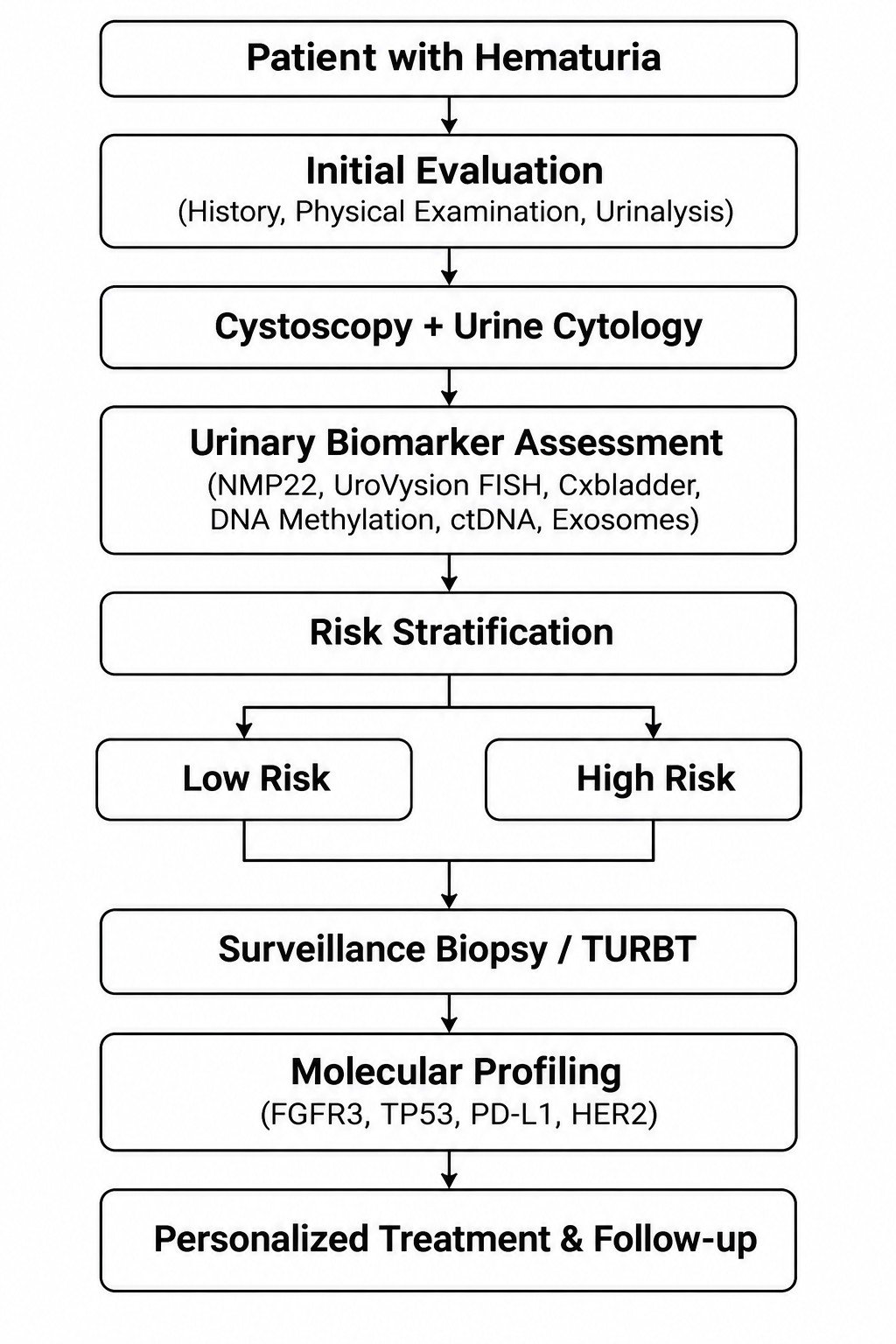
Figure 1: Proposed diagnostic and biomarker-guided management pathway for bladder cancer.
Challenges and Limitations
Despite impressive early results, several barriers remain. First, assay variability and standardization are major issues: different studies use different platforms, cutoffs, and sample types, making comparisons difficult. For example, PD-L1 IHC results vary by antibody clone and scoring method, contributing to its inconsistent predictive value. Similarly, urinary miRNA and methylation studies often lack protocol harmonization, leading to heterogeneity in reported performance. Second, cohort heterogeneity and bias limit generalizability: many studies are single-center, retrospective, or enriched for advanced disease. Small sample sizes and lack of proper controls can inflate accuracy. Third, cost and complexity pose hurdles: next-generation sequencing and exosome isolation are expensive and not widely available. High-throughput assays (NGS panels, methylation arrays) may not be feasible in routine practice without streamlining. Fourth, false positive/negatives are unavoidable: inflammation, infections, or clonal hematopoiesis can produce spurious signals (e.g. low-level DNA mutations in urine). Conversely, low-grade tumors may shed too little material, yielding false negatives. Finally, regulatory and clinical adoption remain challenging. No new urinary or tissue biomarker (beyond FDA-approved test like UroVysion or FDA-cleared assays) has yet been incorporated into guidelines. Demonstrating that a biomarker strategy improves patient outcomes (not just test accuracy) will be essential. In summary markers-driven algorithms must overcome technical variability, validate in diverse cohorts, and demonstrate clear benefit over existing standards.
Future Perspective and Research Priorities
Looking ahead, the field is moving toward integrated, precision diagnostics. Multi-osmics assays that combine DNA mutations, methylation, RNA (mRNA/miRNA), and protein markers from the same urine or blood sample may capture tumor biology more comprehensively. Early studies suggest that integrating genomics (e.g. TERT/FGFR3 mutations), epigenetic, and exosomal markers can substantially increase accuracy. Artificial Intelligence (AI) and machine learning will be critical to make sense of high-dimensional data. Preliminary models using AI to analyze composite biomarkers and imaging data have achieved AUCs >0.90 but need large training sets and external validation. Future trials should incorporate biomarkers-guided arms: for example, randomizing high-risk NMIBC patients (based on a new biomarker index) to intensified therapy vs. standard care. Similarly, “umbrella” trials could direct metastatic patients with FGFR3 mutations to erdafitinib and those with PD-L1 expression to immunotherapy, prospectively testing biomarkers-driven therapy selection. Efforts to standardize protocols are underway (e.g. international consortia and consortia-driven biobanks). Health-economic studies should run in parallel, evaluating cost per avoided cystoscopy or per quality-adjusted life-year gained by biomarker adoption. Notably, updating risk calculators (like EORTC or CUETO models for NMIBC) to include molecular factors could refine individual risk estimates. Overall, the research agenda prioritizes (1) validating promising markers in prospective multicenter trials, (2) developing and validating multi-biomarker panels using robust pipelines, (3) integrating clinical data with molecular classifiers via AI, and (4) establishing regulatory frameworks and reimbursement pathways. International collaboration and data sharing will accelerate progress.
Conclusion
Tissue and urinary biomarkers offer the promise of transforming bladder cancer care beyond the blunt tools of cytology and cystoscopy. Tissue markers like FGFR3, TP53, Ki-67, PD-L1, and HER2 illuminate tumor biology and, in some cases, guide targeted therapy. When combined with urinary DNA/RNA assays, they could enable personalized triage, surveillance, and treatment algorithms. To realize this potential, however, rigorous validation and standardization are needed. Prospective trials should test whether marker-driven strategies truly improve outcomes. In the coming years, we anticipate a shift towards “precision urology” where invasive procedure and one-size fits-all surveillance give way to risk-adjusted biomarker-guided management. As multi-omics technologies mature and cost fall, integrated molecular profiling (possibly aided by AI) may finally allow clinicians to tailor interventions to each patient’s unique tumor profile.
Ethical Approval
Ethical approval was not required for this narrative review.
Data Availability
Data sharing is not applicable to this article as no datasets were generated or analyzed during the current study.
References
- Lenis AT, Lec PM, Chamie K, Mshs MD. Bladder cancer: A review. JAMA. 2020;324(19):1980-91. PubMed ↗
- Sung H, Ferlay J, Siegel RL, Laversanne M, Soerjomataram I, Jemal A, et al. Global cancer statistics 2020: GLOBOCAN estimates of incidence and mortality worldwide for 36 cancers in 185 countries. CA Cancer J Clin. 2021;71(3):209-249. PubMed ↗
- Batista R, Vinagre N, Meireles S, Vinagre J, Prazeres H, Leão R, et al. Biomarkers for bladder cancer diagnosis and surveillance: A comprehensive review. Diagnostics (Basel). 2020;10(1):39. PubMed ↗
- van Rhijn BW, van der Poel HG, van der Kwast TH. Urine markers for bladder cancer surveillance: A systematic review. Eur Urol. 2005;47(6):736-748. PubMed ↗
- Knowles MA, Hurst CD. Molecular biology of bladder cancer: New insights into pathogenesis and clinical diversity. Nat Rev Cancer. 2015;15(1):25-41. PubMed ↗
- Richters A, Aben KKH, Kiemeney LALM. The global burden of urinary bladder cancer: An update. World J Urol. 2020;38(8):1895-1904. PubMed ↗
- Böyük A, Şanlı Ö, Erdem S, Tefik T, Özcan F, Özlük Y, et al. The association between variant urothelial histologies, pathological stage and disease-specific survival in patients with bladder cancer. Turk J Urol. 2018;44(1):24-30. PubMed ↗
- Holzbeierlein JM, Bixler BR, Buckley DI, Chang SS, Holmes R, James AC, et al. Diagnosis and treatment of non-muscle invasive bladder cancer: AUA/SUO Guideline: 2024 Amendment. J Urol. 2024;211(4):533-538. PubMed ↗
- Devlies W, de Jong JJ, Hofmann F, Bruins HM, Zuiverloon TCM, Smith EJ, et al. The diagnostic accuracy of cystoscopy for detecting bladder cancer in adults presenting with hematuria. Eur Urol Focus. 2024;10(1):115-122. PubMed ↗
- Chen F, Lin X. The Paris system for reporting urinary cytology: An updated review. J Clin Transl Pathol. 2023;3(2):59-74. XiahePublishing ↗
- Woo S, Panebianco V, Narumi Y, Del Giudice F, Muglia VF, Takeuchi M, et al. Diagnostic performance of VI-RADS for prediction of muscle-invasive bladder cancer: A systematic review and meta-analysis. Eur Urol Oncol. 2020;3(3):306-315. PubMed ↗
- Koga F. What are roles of multiparametric MRI prior to transurethral resection of bladder tumor? Transl Androl Urol. 2019;8(4):290-291. PMC ↗
- Russo GI, Sholklapper TN, Cocci A, Broggi G, Caltabiano R, Smith AB, et al. Performance of Narrow Band Imaging (NBI) and Photodynamic Diagnosis (PDD). Cancers (Basel). 2021;13(17):4378. PubMed ↗
- Mowatt G, Zhu S, Kilonzo M, Boachie C, Fraser C, Griffiths TR, et al. Systematic review of photodynamic diagnosis and urine biomarkers. Health Technol Assess. 2010;14(4):1-331. PubMed ↗
- Wang Z, Que H, Suo C, Han Z, Tao J, Huang Z, et al. Evaluation of the NMP22 BladderChek test for detecting bladder cancer. Oncotarget. 2017;8(59):100648-100656. PubMed ↗
- O'Sullivan P, Sharples K, Dalphin M, Davidson P, Gilling P, Cambridge L, et al. A multigene urine test for bladder cancer detection. J Urol. 2012;188(3):741-747. PubMed ↗
- Lotan Y, Daneshmand S, Shore N, Black P, Scarpato KR, Patel A, et al. Cxbladder Triage versus cystoscopy. J Urol. 2024;212(1):41-51. PubMed ↗
- Yang Z, Song F, Zhong J. Urinary biomarkers in bladder cancer: FDA-approved tests and emerging tools. Cancers (Basel). 2025;17(21):3425. PubMed ↗
- Silva-Ferreira M, Carvalho JA, Salta S, et al. Diagnostic accuracy of urinary DNA methylation biomarkers. Eur Urol Focus. 2024;10(6):922-934. PubMed ↗
- Witjes JA, Morote J, Cornel EB, et al. Performance of the Bladder EpiCheck™ methylation test. Eur Urol Oncol. 2018;1(4):307-313. PubMed ↗
- Ruan W, Chen X, Huang M, et al. A urine-based DNA methylation assay. Clin Epigenetics. 2021;13(1):91. PubMed ↗
- Xiao S, Wang J, Xiao N. MicroRNAs as noninvasive biomarkers. Int J Biol Markers. 2016;31(3):e276-e285. PubMed ↗
- Long C, Shi H, Li J, et al. Diagnostic accuracy of urine-derived exosomes. World J Surg Oncol. 2024;22(1):285. PMC ↗
- Dudley JC, Schroers-Martin J, Lazzareschi DV, et al. Detection and surveillance using urine tumor DNA. Cancer Discov. 2019;9(4):500-509. PubMed ↗
- Chauhan PS, Shiang A, Alahi I, et al. Urine cell-free DNA multi-omics. NPJ Precis Oncol. 2023;7(1):6. PubMed ↗
- Dudderidge T, Stockley J, Nabi G, et al. ADXBLADDER performance evaluation. Eur Urol Oncol. 2020;3(1):42-46. PubMed ↗
- Harsanyi S, Kianickova K, Katrlik J, et al. Proteomic and glycomic biomarkers. J Cancer Res Clin Oncol. 2024;150(2):96. PubMed ↗
- Wang R, Kang H, Zhang X, et al. Urinary metabolomics by UPLC-MS. BMC Cancer. 2022;22(1):214. PubMed ↗
- Meng XY, Zhou XH, Li S, et al. Machine learning-based detection using urine cfDNA fragmentation hotspots. Clin Chem. 2024;70(12):1463-1473. PubMed ↗
- Laurie MA, Zhou SR, Islam MT, et al. Bladder cancer and artificial intelligence. Urol Clin North Am. 2024;51(1):63-75. PubMed ↗
- Fong MHY, Feng M, McConkey DJ, Choi W. Update on bladder cancer molecular subtypes. Transl Androl Urol. 2020;9(6):2881-2889. PubMed ↗
- Cathomas R, Lorch A, Bruins HM, et al. 2021 EAU guidelines on metastatic urothelial carcinoma. Eur Urol. 2022;81(1):95-103. PubMed ↗
- Missaoui N, Bdioui A, Baccouche A, et al. Significance of p53, p27, Ki-67, E-cadherin and HER2 expression. J Egypt Natl Canc Inst. 2020;32(1):36. PubMed ↗