Birthmarks: Typical and Atypical Presentations - Case Series
Abstract
This article describes birthmarks and their clinical approach in the context of pediatric outpatient care. The aim is to raise awareness among physicians about these medical conditions and optimize their clinical approach in primary care.
Methodology: A descriptive study of a cohort of 12 children under ten years of age who were diagnosed with a birthmark between August 2022 and Augst 2025 in a pediatric primary care and family medicine center in Huttwil, Switzerland was conducted.
Discussion: Birthmarks are common and generally benign; however, some require early identification in primary care for proper follow-up and treatment.
Keywords
Birthmark; Cutaneous lesions in Newborns; Pediatric primary care
Introduction
Birthmarks are benign skin lesions present at or shortly after birth. They are estimated to affect 10% to 50% of children. The incidence is influenced by some factors such as race and ethnicity [1]. Most birthmarks are harmless and require no treatment, while others, such as hemangiomas, have recently benefited from new treatment and follow-up strategies.
Some authors have evaluated the management and diagnosis of birthmarks by healthcare professionals, identifying diagnostic errors and/or difficulties managing the lesions [2,3].
The definition of the birthmark categories will be followed by a description of some of the most relevant clinical and epidemiological data for each category. Finally, we will present an overview of the patient cohort that was studied.
Theoretical Background
There are three categories of birthmarks:
i. Pigmented birthmarks: Are due to increased melanocytes, melanin deposition, or congenital melanocytes or melanin distribution (Table 1).Table 1: Pigmented birthmark [4].
| Type | Epidemiological Points | Clinical Findings | Diagnostic Assessment | Management Principles |
|---|---|---|---|---|
| Dermal melanocytosis (Mongolian spots) [5] | Frequently found in children with dark skin. An association has been found between the presence of extensive Mongolian spots in atypical locations and underlying pediatric lysosomal storage diseases [4]. | Congenital skin discoloration can be singular or multiple. Such manifestations are usually located in the lumbosacral region and are seldom encountered in the extremities, buttocks, shoulder, or trunk. | Clinical observation. The disappearance during infancy and the extremely rare persistence of the condition into adulthood are both features of the condition. | Photographic documentation. A possible differential diagnosis is a hematoma due to physical abuse. |
| Pigmented nevi – moles [6] | Present at birth or during the first week. Different sizes are available. The sizes are small (less than 1.5 cm), medium-sized (1.5–20 cm), and large/giant (more than 20 cm). | Initially flat and tan. Later, they lighten or darken. They eventually develop nodules, satellite lesions, or areas of hypertrichosis. | Clinical observation. Large nevi are associated with a higher risk of melanoma transformation. |
Monitoring using the melanoma guide for children:
|
| Café-au-lait spots [7] | Common. The number and distribution may indicate potential genetic disease. |
Smooth, well-demarcated macules ranging in color from light brown to dark brown.
Associated potential genetic diseases include:
|
Clinical observation. Six macules greater than 0.5 cm in size may indicate a suspected associated genetic syndrome. | Referral for early screening of genetic diseases. |
Table 2: Vascular Malformations [8,9].
| Types | Epidemiological Points | Clinical Findings | Diagnostic Assessment | Management Principles |
|---|---|---|---|---|
| Hemangioma |
Congenital hemangiomas. Infantile hemangioma. Multiple hemangiomas those in special regions (head, neck and anogenital region). |
Present at birth. They may involve spontaneously (RICH rapidly involving
congenital hemangioma), or they may not involute (NICH: non-involving
congenital hemangioma), or they may show partial involution (PICH: Partially
involuting congenital hemangioma). They appear in the first weeks. They grow rapidly and then involute. Multifocal infantile hemangiomas: Five hemangiomas suspected to be diffuse neonatal hemangiomatosis with hepatic involvement. Segmental hemangioma in the facial region is associated with PHACES syndrome. Multiple or large hemangioma on the lower body is a sign of LUMBAR syndrome. Large hemangiomas may be associated with cardiac and hypothyroidism. |
Clinical observation. Identify hemangiomas that require treatment. Life-threatening complications, permanent disfigurement or functional impairment. Abdominal ultrasound. Echocardiography - MRI of the head. Spine and Kidneys Ultrasound. |
Topical or oral propranolol. The timing of treatment initiation is important. |
| Malformation capillary |
Salmon patches/ stork bites / Angel kiss. Port wine stain/ Nevus flammeus. Cutis marmorata telangiectatica congenita. |
Typically, the location is along the midline. Unilateral pink or discoloration is common. These lesions are most frequently observed on the face and neck. Differential diagnosis: Cutis marmorata physiologica; The marks do not fade with warming. |
Clinical examination. Ultrasound of slow-flow malformation. In the first-year life, most lesions usually fade to some extent, but patches that are slightly darker in color than the surrounding skin can sometimes be left behind. These patches can also leave a mark on the skin. |
No treatment is required. Disappears over time. It persists throughout life. No treatment is required. Laser. |
| Lymphatic malformations |
Microcystic macrocystic mixed. 90% of case occurs within the first two years. |
Voluminous subcutaneous masses often develop in the head and neck area.
Blood-filled vesicles may also appear on the skin or in the mouth. These
masses grow alongside the child. Congenital truncal lipomatous overgrowth. |
Clinical examination - require correct classification. | Referral. |
| Venous Malformation |
Can be either solitary or multifocal. Associated extracutaneous localizations; most frequently on the gastrointestinal tract. CLOVES Syndrome. |
Lymphatic malformation. Epidermal nevi. Scoliosis and spinal anomalies. Compressible, appear blue, soft and often deep. They grow with child. Congenital truncal lipomatous overgrowth. Vascular malformation. Epidermal nevi. Scoliosis and spinal anomalies. |
Table 3: Other lesions [10,11].
| Type | Epidemiological Points | Clinical Findings | Diagnostic Assessment | Management Principles |
|---|---|---|---|---|
| Dermal melanocytosis (Mongolian spots) [5] | Frequently found in children with dark skin. An association has been found between the presence of extensive Mongolian spots in atypical locations and underlying pediatric lysosomal storage diseases [4]. | Congenital skin discoloration can be singular or multiple. Such manifestations are usually located in the lumbosacral region and are seldom encountered in the extremities, buttocks, shoulder, or trunk. | Clinical observation. The disappearance during infancy and the extremely rare persistence of the condition into adulthood are both features of the condition. | Photographic documentation. A possible differential diagnosis is a hematoma due to physical abuse. |
| Pigmented nevi – moles [6] | Present at birth or during the first week. Different sizes are available. The sizes are small (less than 1.5 cm), medium-sized (1.5–20 cm), and large/giant (more than 20 cm). | Initially flat and tan. Later, they lighten or darken. They eventually develop nodules, satellite lesions, or areas of hypertrichosis. | Clinical observation. Large nevi are associated with a higher risk of melanoma transformation. |
Monitoring using the melanoma guide for children:
|
| Café-au-lait spots [7] | Common. The number and distribution may indicate potential genetic disease. |
Smooth, well-demarcated macules ranging in color from light brown to
dark brown.
Associated potential genetic diseases include:
|
Clinical observation. Six macules greater than 0.5 cm in size may indicate a suspected associated genetic syndrome. | Referral for early screening of genetic diseases. |
Patient Information
A retrospective descriptive study of a cohort of twelve children under ten years of age, diagnosed with a birthmark between August 2022 and August 2025, was conducted at a pediatric primary care and family medicine center in Huttwil, Switzerland.
Data were extracted from electronic medical records, including demographic information, clinical characteristics, diagnostic assessments and treatment outcomes.
Most were diagnosed with birthmarks during well-child Check-ups; some patients were diagnosed during unscheduled visits for acute illness unrelated to birthmarks (Patient 1-12).
Discussion
Birthmarks are a common finding during medical examinations of newborns and children. Symptoms are usually mild and may include discharge, minor anatomical abnormalities, increased pigmentation or hair growth in unusual areas.
Clinical management requires early and accurate diagnosis to avoid over treatment and/or to ensure pediatric patients benefit from current treatments, such as local propranolol, laser therapy, or referral, to prevent complications from delayed diagnosis.
Some key facts about each type of birthmark have been summarized to facilitate its clinical management.
Acknowledgement
We would like to thank the young patient and his family.
Ethics statement
Ethical approval was not required.
Declaration of Competing Interests
The Author declares no competing interests.
Informed consent
Is not required. The information is based on internal clinical studies of medical history records from the practice, with patient data kept anonymous.
References
- Kanada KN, Merin MR, Munden A, Friedlander SF. A prospective study of cutaneous findings in newborns in the United States: Correlation with race, ethnicity, and gestational status using updated classification and nomenclature. J Pediatr. 2012;161(2):240-5. PubMed
- Rayala BZ, Morrell DS. Common skin conditions in children: Neonatal skin lesions. FP Essent. 2017;453:11-7. PubMed
- Berthelot B, Prémel J, Pichereau M, Moulinier E, Vabres P, Martin L. How congenital skin lesions are perceived by health professionals in the delivery room-the reconnaissance study. Pediatr Dermatol. 2026;43(1):43-8. PubMed
- Bartenstein Reusch D, Hawryluk EB. Pigmented lesions in children: Update on clinical, histopathologic and ancillary testing. Dermatol Clin. 2022;40:25-36. PubMed
- Athanti S, Mouzam S, Sohail Ahmed M. Atypical Mongolian spots with hurler's disease: A case report. Cureus. 2024;16(4):e58501. PubMed
- Mologousis MA, Tsai SYC, Tissera KA, Levin YS, Hawryluk EB. Updates in the management of congenital melanocytic nevi. Children. 2024;11(1):62. PubMed
- Albaghdadi M, Berseneva M, Pennal A, Wan S, Matviychuk D, Shugar A, et al. Value of a café-au-lait macules screening clinic: Experience from the hospital for sick children in Toronto. Pediatr Dermatol. 2022;39(2):205-10. PubMed
- Seront E, Hermans C, Boon L, Vikkula M. Targeted treatments for vascular malformations: Current state of the art. J Thromb Haemost. 2024;22(11):2961-75. PubMed
- Léauté-Labrèze C, Baselga Torres E, Weibel L, Boon LM, El Hachem M, van der Vleuten C, et al. The infantile hemangioma referral score: A validated tool for physicians. Pediatrics. 2020;145(4):e20191628. PubMed
- Brown KW, Lucas E, Hoppe IC, Humphries LS. A review on lumps, bumps, and birthmarks: When and why to refer. Pediatr Ann. 2023;52(1):e23-30. PubMed
- Bessis D, Société française de dermatologie pédiatrique. [Cutaneous signs of occult cranial and spinal dysraphism]. Ann Dermatol Venereol. 2020;147(8-9):504-19. PubMed
Patient 1-2: Pigmented birthmark.
| Types | Epidemiological points | Clinical Findings | Diagnostic Assessment | Management Principes |
|---|---|---|---|---|
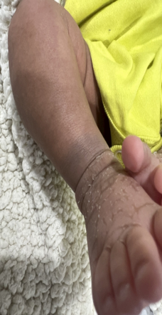
| Patient 1: Dermal melanocytes (Mongolian spots) | A one-month -old female has had a single dark blue mark on her tibiotarsal joint since birth. |
|
The Mongolian spot is found in an unusual location, with 0.98% of case occurring on the extremities. | Photographic documentation. |
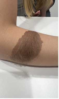
| Patient 2: Pigmented nevi (mola) | A 10- year-Old girl who has had a hyperpigmented patch on her right forearm since birth. |
|
A pigmented naevus that has developed an area of hypertrichosis. | Clinical observation |
Patient 3-5: Vascular malformations.
| Types | Epidemiology Clinical classification | Clinical Findings | Diagnostic Assessment | Therapeutic intervention |
|---|---|---|---|---|
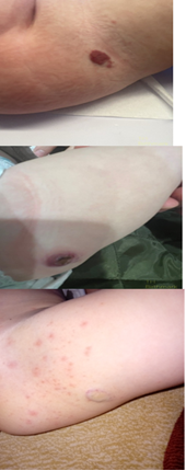
| Patient 3: Hemangioma | A girl, aged six months, had a 2 cm hemangioma in the right medial femoral region. |
|
By the time she was nine months old, the hemangioma had ulcerated
(diaper). The hemangioma regressed Spontaneously at the aged of three. |
Clinical observation. |
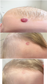
| Patient 4: Hemangioma | A one-month-old girl hat a hemangioma in the frontal region. |
|
Photographic follow-ups are to be carried out at two months, six months and nine months. | Topical propanol. |
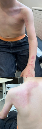
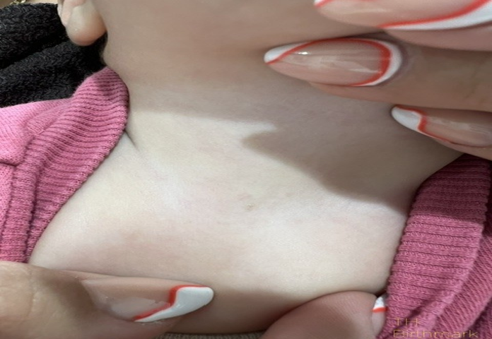
| Patient 5: Port wine stain/ Nevus flammeus. | A six- year- old boy has had nevus flammeus on his arm, chest and neck. Since birth. |
|
Reduction in pigmentation during the clinical course and during laser therapy. | Laser |
Patient 6-12: Other lesions; Fibrous hamartoma, branchial sinuses, ski markers of occult spinal dysraphism, congenital dermoid fistulas of the anterior chest region.
| Types | Epidemiology Clinical classification | Clinical Findings |
|---|---|---|
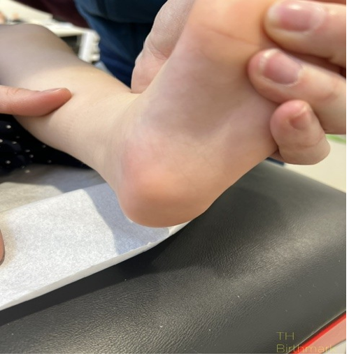
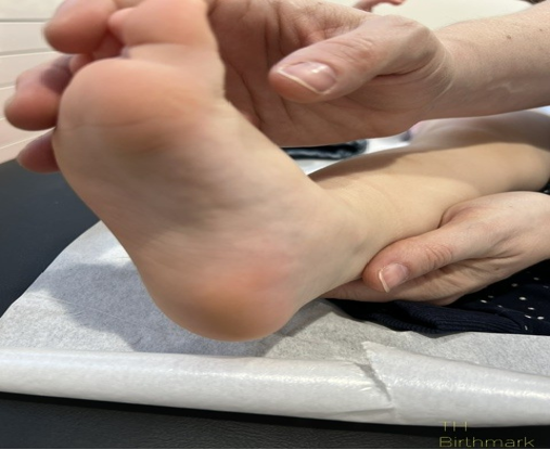
| Patient 6: Precalcaneal congenital fibrolipomatous hamartoma bilateral |
A nodule is present Gird’s foot. It is bilateral and located in the
calcaneal region. Her mother first noticed it on the second day of her life. |
|
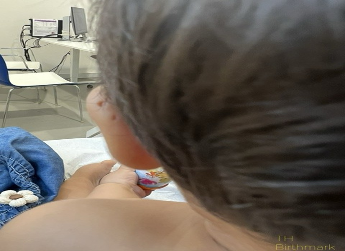
| Patient 7: First branchial cleft anomalies | A nine-month-old girl present with a unilateral auricular fossa, has been present since birth. |
|
| Patient 8: Sacral Dimples. | A two-month-old girl has a dimple that is 1.5 cm from the anal orifice and 0.5 centimeters in diameter. |
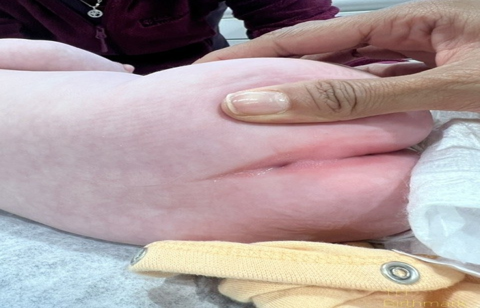
|
| Patient 9: Sacral Dimples. | A one-month-old girl has a dimple that is 3 cm from the anal orifice and 1 centimeter in diameter. |
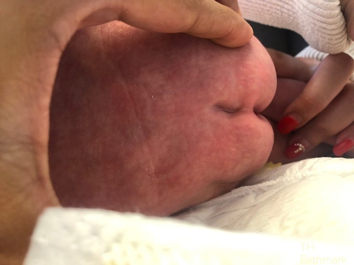
|
| Patient 10: Aplasia Cutis | A four-month-old girl with a scalp spot measuring 2 cm by 2 centimeters. |
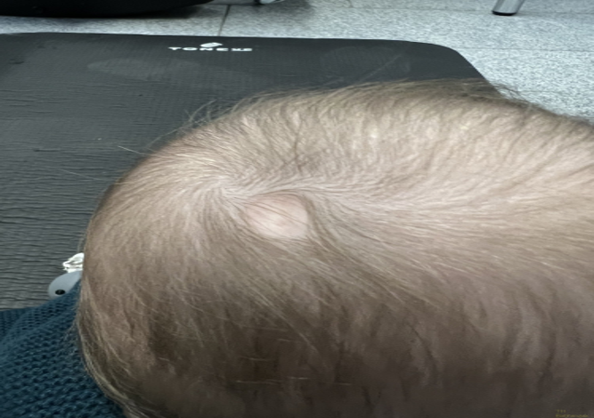
|
| Patient 11: Congenital dermoid fistulas of the anterior chest region. | A 14-months-old boy hat a small congenital dimple on the manubrium of the sternum. |
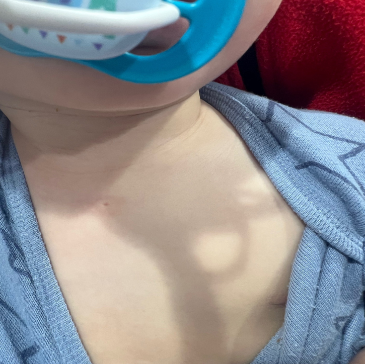
|
| Patient 12: Congenital dermoid fistulas of the anterior chest region. | A nine-month- old girl has a fistula with intermittent discharge in the sternal region. |
|