Targeting Metabolism and Disulfidptosis: A Novel strategy for Cancer Therapy
Abstract
Disulfidptosis represents a recently characterized form of programmed cell death driven by an intracellular imbalance between cystine and the vital reductant Nicotinamide Adenine Dinucleotide Phosphate (NADPH). This metabolic perturbation precipitates an excessive accumulation of disulfide bonds within cytoskeletal proteins, ultimately compromising cellular integrity and inducing death. Specifically, under conditions of glucose starvation, the restricted supply of intracellular NADPH fails to reduce the aberrant influx of cystine, thereby triggering the disulfidptotic cascade. This review provides a comprehensive critical synthesis of the molecular mechanisms underlying disulfidptosis, with a particular focus on the pivotal roles of solute carrier family 7 member 11 (SLC7A11) and the Rac1-WAVE Regulatory Complex (WRC) signaling axis. Beyond delineating the mechanistic framework, we contextualize these findings within the broader landscape of translational cancer therapy. We systematically evaluate emerging therapeutic strategies designed to exploit this metabolic vulnerability, including the targeted inhibition of SLC7A11 and the pharmacological modulation of glucose, cystine, and NADPH metabolism (via nicotinamide adenine dinucleotide kinase and the pentose phosphate pathway). Furthermore, we discuss the complex interplay between disulfidptosis, tumor metabolic reprogramming, and the immunosuppressive tumor microenvironment. By critically analyzing these interconnected pathways, this review aims to provide conceptual insights and highlight novel, combinatorial therapeutic avenues for precision oncology.
Keywords
Cell metabolism; Tumor therapy; Disulfidptosis; Programmed cell death; Solute carrier family
Introduction
Disulfidptosis has recently emerged as a distinct, metabolic-stress-induced modality of regulated cell death. Its defining feature is the severe perturbation of the intracellular homeostatic balance between cystine and the critical reducing agent Nicotinamide Adenine Dinucleotide Phosphate (NADPH). When this delicate redox equilibrium is disrupted, cells experience an overwhelming oxidation of protein sulfhydryl groups. This pathological shift drives the aberrant formation of intra- and intermolecular disulfide linkages, predominantly within actin cytoskeletal proteins, thereby fatally compromising cellular architecture and function [1,2] (Figure 1). Furthermore, studies indicate that under glucose starvation, cells overexpressing the cystine/glutamate antiporter (xCT) demonstrate significantly enhanced L-cystin uptake, a phenomenon particularly observed in renal cancer cells [3]. The biological production of glutathione (GSH), a paramount endogenous antioxidant providing cellular defense from injurious oxidation, is critically reliant upon cystine, which serves as its essential foundational molecule. However, the reduction of cystine consumes NADPH. Under glucose starvation, restricted intracellular NADPH generation leads to the accumulation of considerable volumes of cytosolic oxidized cystine. Excess intracellular cystine then facilitates the generation of aberrant disulfide bonds within and between protein molecules, thereby disrupting their normal structure and function and ultimately causing cell death.
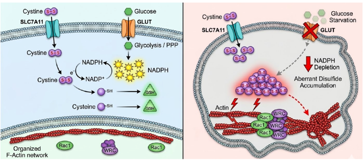
Figure 1: Molecular mechanism of disulfidptosis: Disulfidptosis is triggered by the disruption of the dynamic balance between cystine and NADPH, leading to excessive formation of disulfide bonds. This process involves regulation of the Rac1-WAVE Regulatory Complex (WRC) signaling pathway and perturbation of cellular redox homeostasis, ultimately resulting in cell death.
Molecular Mechanism of Disulfidptosis
Solute carrier family 7 member 11 (SLC7A11)The processes of cellular proliferation, metabolic process, and ultimate demise are fundamentally influenced by the protein SLC7A11. This essential conveyer of amino acids exerts such critical control through its capacity to alter the internal amino acid profile in conjunction with the overall redox condition [4,5]. By primarily regulating intracellular cystine levels, SLC7A11 influences the cellular redox state and thereby contributes to the modulation of disulfidptosis. SLC7A11 transports cystine into cells, providing the substrate for glutathione (GSH) synthesis. Excessive cystine during oxidative stress triggers disulfide bond formation which results in protein inactivation and cellular dysfunction [6]. NADPH functions as a vital intracellular reductant which gets used up during cystine reduction. SLC7A11 overexpression leads to NADPH depletion which intensifies oxidative stress and triggers disulfidptosis [7].
Transcription factors ATF4, Nrf2, and p53 regulate the expression of the SLC7A11 gene [4,8]. Furthermore, SLC7A11 influences multiple biological processes including cellular metabolism as well as the mechanisms of ferroptosis and autophagy [9]. The upregulation of SLC7A11 expression results in reduced glutamate levels inside cells which makes cells more reliant on glutamine [10]. Furthermore, SLC7A11 affects ferroptosis regulation through GSH synthesis modulation and GPX4 activation which boosts cellular antioxidant capacity [11]. Additionally, the absence of SLC7A11 triggers autophagy through LC3-II accumulation [12]. The amino acid transporter SLC7A11 demonstrates multiple effects on cell growth, metabolism and cell death. SLC7A11 influences multiple cell death pathways through its control of intracellular amino acid levels and redox balance including disulfidptosis [13]. Extensive research into the functions of SLC7A11 remains essential to advance our knowledge of tumor development and treatment.
The Rac1-WRC pathwayA principal contribution of the Rac1-WAVE Regulatory Complex (WRC) signaling axis lies in the remodeling of the cytoskeleton, with a particular emphasis on governing the dynamic behavior pertaining to actin-based filaments [6]. As an integral component of the Rho GTPase family, Rac1 fulfills the multiple functions of cell biology [14]. Rac1 exerts substantial influence across a wide spectrum of cellular operations. These encompass, among others, alterations to the internal cellular architecture, mechanisms of intercellular bonding, directed cellular movement, the capacity for tissue infiltration, generation of oxidative molecules, engagement in immune system activation, the trafficking of membrane-bound compartments, development of new vasculature, processes of cellular multiplication, and pathways leading to programmed cellular demise [15-17]. Furthermore, the protein Rac1 is understood to participate in disulfidptosis - a recently identified mechanism for programmed cellular elimination-through a process reliant on WRC protein action that culminates in the assembly of branched actin structures [18]. Therefore, the communicative molecular cascade involving both Rac1 and the aforementioned WRC is considered to hold a decisive modulatory capacity over the disulfidptotic event. Mechanistically, the WRC complex maintains cytoskeletal stability by modulating actin dynamics. Subsequent to an initial association, Rac1 elicits stimulation within the WRC; this critical process involves interaction with the WRC's distinct WCA element, a domain incorporating WASP-related homology type 2 (WH2) plus Central-Acidic (CA) segments. Such specific recruitment of the WCA element thereafter serves to propel actin filament assembly, a mechanism reliant on the Arp2/3 protein machinery [19], culminating in the generation of broad, F-actin-dense membrane protrusions termed lamellipodia. The formation of these structures is hypothesized to enhance intracellular disulfide bond formation, thereby triggering disulfidptosis.
Tumor Therapeutic Strategies Based on Disulfidptosis
Fundamental to understanding disulfidptosis-a recently identified modality of malignant cell self-destruction-is the significant derangement observed in the homeostatic control pertaining to thiol-dependent oxidative states within the cell [2]. To maintain the reducing environment required for malignant growth, tumor cells often upregulate the cystine/glutamate antiporter SLC7A11. However, this high dependence on cystine renders tumor cells highly sensitive to metabolic stress. Upon encountering adverse conditions, for instance glucose scarcity, neoplastic cells undergo a swift decline in their internal reserves of reduction potential. This pronounced diminishment precipitates an overabundance of proteins in an oxidized state, a situation that inexorably culminates in cell death.
Targeting SLC7A11Functioning as an essential component within the disulfidptotic pathway, an overabundance of the SLC7A11 protein exhibits a profound correlation with heightened neoplastic severity, pronounced infiltrative capacity, plus a diminished susceptibility toward established medical interventions. Therefore, it might act as a biomarker for predicting tumor sensitivity to disulfidptosis [20]. Targeting SLC7A11 or interfering with cystine metabolism not only enables the selective killing of tumor cells but also offers novel strategies for overcoming tumor drug resistance [21-23]. SLC7A11 is an established therapeutic target in oncology and a key protein regulating ferroptosis. Various SLC7A11 inhibitors, including Erastin, Imidazole Ketone Erastin (IKE), Sulfasalazine, and Sorafenib, are widely employed in both research and clinical settings. Yet, when considering SLC7A11 as a target for anti-cancer strategies, antecedent work has generally given principal attention to initiating ferroptotic cell death. The approach typically involved preventing the internalization of cystine, a process dependent upon SLC7A11 activity [24]. Consequently, these agents are often referred to as ferroptosis inducers. Erastin is one of the most widely used SLC7A11 inhibitors and ferroptosis inducers in cell culture studies [25]. IKE, an Erastin analog, demonstrates inhibitory activity against various tumors, including lung cancer, pancreatic cancer, lymphoma, and melanoma [26-28]. The pharmaceutical agents Sorafenib, along with Sulfasalazine, possess official sanction from the United States entity responsible for oversight concerning foodstuffs plus medicinal agents (FDA). Serving as multi-kinase inhibitor, Sorafenib has demonstrated significant efficacy in treating various tumors, including renal cancer and hepatocellular carcinoma [29,30]. Sulfasalazine, which inhibits prostaglandin synthesis, is commonly used to treat patients with Inflammatory Bowel Disease (IBD) [31]. These two compounds share a common mechanistic profile, which includes curtailing the functional capacity of the SLC7A1 transporter. Furthermore, they promote the onset of ferroptotic cell demise and concurrently attenuate neoplastic expansion. However, the strategy of targeting SLC7A11 to inhibit tumor cell proliferation via disulfidptosis remains underexplored. Further investigation in this area could yield novel therapeutic strategies and perspectives for cancer treatment.
Glucose metabolismInhibiting glucose transport represents a potential therapeutic strategy [32]. By blocking glucose uptake, glucose transport inhibitors reduce intracellular NADPH levels, thereby inducing disulfidptosis in tumor cells. Inhibitors targeting Glucose Transporters (GLUTs) have demonstrated anticancer efficacy. Notably, the GLUT inhibitor BAY-876 effectively inhibits glucose uptake in tumor cells. BAY-876 exhibits long-lasting antitumor activity, inhibiting glycolysis and proliferation in hepatocellular carcinoma. Besides, in triple-negative breast cancer, it demonstrates greater cytotoxicity towards cells characterized by high glycolysis and low oxidative phosphorylation rates [33-35]. As a direct repercussion, such curtailment results in lessened NADPH generation and, in parallel, a significant upsurge within the NADP+ vs. NADPH quotient. As a direct outcome, such a scenario fosters the irregular creation of disulfide linkages among protein constituents forming the cell's actin framework, alongside a structural failure impacting filamentous actin assemblages. This conjunction of disruptive events subsequently leads to the initiation of disulfidptotic programmed cell death.
Cystine metabolismThe amino acid cystine holds critical importance regarding the upkeep of intracellular redox equilibrium. Furthermore, it functions like a vital building block for prominent antioxidant molecules; glutathione exemplifies this [36-38]. Given that a pro-oxidant state inherently characterizes the milieu found external to cellular confines, cysteine is rapidly oxidized to cystine extracellularly, resulting in significantly lower extracellular cysteine concentrations relative to cystine [39]. Therefore, a substantial proportion of cellular entities demonstrate a strong reliance upon the SLC7A11 mechanism for obtaining cystine from the external environment to satisfy inherent needs. This widespread dependence, in turn, curtails the scope of approaches designed to trigger disulfidptotic demise by altering the accessibility of cystine.
Functioning as a crucial antioxidative apparatus within cellular confines is the Thioredoxin (Trx) assembly. Its principal elements encompass the Trx protein itself, an enzyme acting as a reductase for Thioredoxin (TrxR), together with NADPH [40]. Within this system, Trx catalyzes the cleavage of protein disulfide bonds. The TrxR catalytically employs NADPH to supply reducing equivalents. These are subsequently used for the Trx from its oxidized condition, a process indispensable for upholding the operational efficacy inherent to the entire Trx pathway [41]. The Trx network is indispensable for the reduction of cystine. Reduced Trx can reduce cystine to cysteine, thereby supplying this essential amino acid for protein synthesis. However, under elevated intracellular oxidative stress, the Trx system becomes overwhelmed and cannot efficiently scavenge excess Reactive Oxygen Species (ROS). This leads to excessive protein oxidation and the accumulation of protein disulfide bonds. The accumulation of these disulfide bonds disrupts normal cellular function, ultimately culminating in cell death [42].
NADPH metabolismNicotinamide Adenine Dinucleotide Kinase (NADK): NADPH, an essential intracellular electron donor, is critical for maintaining redox homeostasis, biosynthesis, and protection against oxidative stress [43,44]. NADK, the key enzyme in NADPH synthesis, regulates intracellular NADPH levels by catalyzing the transformation of NAD+ to NADP+ [45]. NADK enables pancreatic cancer cells to overcome hypoxic stress and maintain proliferation [46]. Furthermore, NADK is instrumental in modulating lipid processing and oxidative stress responses during breast cancer metastasis [47]. Similarly, inhibiting NADK activity reduces intracellular NADPH levels, prompting a decline in antioxidant quantities such as glutathione, thereby inducing oxidative stress. This accumulation of oxidative stress brings about excessive protein oxidation and extensive protein disulfide bond formation. This disruption of protein structure and function ultimately induces cell death, a process termed disulfidptosis. Targeting NADK represents an emerging strategy for cancer therapy, given that tumor cells typically exhibit high metabolic demands and consequently have an increased requirement for NADPH. Therefore, inhibiting NADPH synthesis in tumor cells allows for their selective killing [48]. Currently, several NADK inhibitors have been developed and demonstrated promising antitumor activity in vitro [49,50]. Nevertheless, deployment in clinical settings pertaining to NADK-blocking compounds currently resides within initial phases. This situation therefore calls for supplementary exploration, aimed at fully ascertaining their therapeutic utility as well as their overall risk profile.
Pentose Phosphate Pathway (PPP): In addition to NADK, other intracellular enzymes and pathways regulate NADPH levels. The PPP, one of the major intracellular metabolic pathways, primarily functions to generate NADPH and ribonucleotide precursors [51]. Operating principally within cellular boundaries as an essential provider of electrons, NADPH assumes a pivotal role across innumerable constructive biologic syntheses. Prominent examples include the metabolic processes dedicated to generating both elongated fatty acyls and sterol compounds, for instance, cholesterol. Thus, this molecule functions like an indispensable enzymatic partner in preserving the reduced form of glutathione within cellular environments; this activity constitutes a pivotal contribution towards safeguarding cells from detrimental oxidative phenomena. Separately, regarding ribonucleotide molecules, which act as foundational building blocks in the creation of nucleic acids, their presence is fundamentally crucial to enable cellular expansion alongside multiplication processes. The rapid proliferation of tumor cells necessitates extensive macromolecular synthesis, thereby significantly increasing the demand for NADPH and ribonucleotides. The PPP is highly active in cancerous cells. By upregulating the abundance of key PPP enzymes, malignant cells can satisfy their elevated requirements for NADPH and ribonucleotides [52]. Furthermore, NADPH generated by the PPP helps maintain the intracellular reducing environment in tumor cells, protecting them from oxidative damage and thereby promoting tumor cell survival and proliferation [53].
Acknowledging a critical bioenergetic route's substantial importance for neoplastic cell viability, the concept of aiming therapeutic interventions at such a metabolic sequence is now viewed as an innovative treatment modality to combat malignant disease. By inhibiting key enzymes of the PPP, such as Glucose-6-Phosphate Dehydrogenase (G6PD) and 6-Phosphogluconate Dehydrogenase (6PGD), intracellular NADPH levels in tumor cells can be reduced. This reduction induces oxidative stress, thereby inhibiting tumor cell growth and proliferation. Compared to normal tissues, the activity of G6PD and 6PGD is increased in various cancers, encompassing gastric, bladder, breast, cervical, and prostate cancer, correlating with poor prognosis and serving as a cornerstone in tumor progression and chemotherapy refractoriness [54-57]. Deficiency of G6PD or 6PGD significantly reduces NADPH levels and enhances chemotherapy-induced apoptosis. Moreover, downregulation of G6PD promotes glucose restriction-induced cell death, whereas its overexpression alleviates this effect. These findings suggest that targeting G6PD and 6PGD could potentially induce disulfidptosis by reducing NADPH levels [58]. G6PD antagonists represent a promising class of clinical therapeutic agents. Dehydroepiandrosterone (DHEA), 6-aminonicotinamide, and G6PDi-1 have been shown to inhibit NADPH production and induce cell death in diseases including lung and breast cancer [59-63].
Targeting the Rac1-WRC axisWhile the modulation of SLC7A11 and metabolic pathways offers direct avenues for inducing disulfidptosis, the translation of the Rac1-WRC signaling axis into a viable therapeutic strategy remains comparatively underdeveloped. As outlined in Section 1.2, Rac1 and the WRC are indispensable for the rapid cytoskeletal remodeling and actin filament branching that physically manifest disulfidptotic cell death. Consequently, pharmacological intervention targeting this axis presents a compelling, albeit challenging, theoretical proposition.
Currently, several small-molecule Rac1 inhibitors, such as NSC23766, EHop-016, and 1A-116, have been developed and evaluated primarily for their capacity to suppress tumor metastasis, invasion, and proliferation. However, their specific application as intentional inducers of disulfidptosis is still in its infancy. At present, leveraging Rac1 inhibitors specifically to trigger or sensitize tumors to disulfide stress remains largely theoretical.
A primary limitation impeding the immediate clinical translation of Rac1-targeted therapies is the ubiquitous physiological role of Rac1 in healthy tissues, including immune cell motility and cardiovascular maintenance. Systemic inhibition poses a significant risk of off-target toxicity. Nevertheless, integrating Rac1-WRC modulation into the disulfidptosis paradigm holds immense future promise. A rational future direction involves the development of tumor-targeted delivery systems (such as antibody-drug conjugates or nanoparticle formulations) for Rac1 modulators, or combining sub-lethal doses of Rac1 inhibitors with glucose metabolism antagonists. By explicitly mapping the intersection of Rac1-mediated cytoskeletal dynamics and metabolic vulnerability, future research can transition this theoretical concept into a synergistic therapeutic reality.
Disulfidptosis and Tumor Metabolism
By means of a profound alteration in their metabolic operations, notably entailing an amplified consumption regarding essential substrates-for instance, the carbohydrate glucose plus the nitrogen-donating amino acid glutamine-neoplastic cellular entities actively cultivate a Tumor Microenvironment (TME) that is a localized niche surrounding the malignancy. This particular intratumoral setting subsequently fosters an expansion of the malignant mass while concurrently impeding host immunological surveillance [9,10]. Such profound metabolic reconfigurations serve a twofold purpose: they furnish the essential energetic fuel required for accelerated neoplastic cell expansion, and concurrently, they diminish the cancer-fighting capabilities of immune system elements. This latter impairment is achieved via modulation of particular physicochemical parameters within the TME, including variations in acidity plus the oxidative state [64]. Moreover, disulfidptotic demise, representing a newly characterized pathway of cellular self-destruction, involves fundamental initiation processes that are deeply intertwined with the distinctive metabolic operations inherent to malignant tissues [65]. This suggests a strong alignment between the metabolic vulnerabilities of tumors and the molecular pathways triggering disulfidptosis. Therapeutic strategies targeting tumor metabolism can influence the tumor immune microenvironment. On one hand, targeting the metabolic vulnerabilities of tumor cells, such as inhibiting glucose transport or glutamine metabolism, can restrict tumor cell growth and foster a more favorable environment for immune cells [66,67]. Conversely, triggering disulfidptotic demise through manipulation of the internal cellular redox equilibrium may perturb protein management mechanisms within neoplastic entities. This subsequently heightens their vulnerability towards identification plus elimination via host immunological surveillance [68]. Furthermore, an over-consumption pertaining to certain amino acid molecules, for instance cystine, by these malignant cells culminates in a scarcity regarding said nutritional elements inside the TME. Such deprivation consequently constrains the expansion along with operational capacity exhibited by immune system components, including T lymphocytes [69-71]. Furthermore, metabolic byproducts generated by tumor cells, such as lactate, can acidify the TME and inhibit T cell activity [72]. The immunosuppressive effects of tumor metabolism within the TME can potentially be countered by targeting disulfidptosis and specifically by targeting cystine metabolism to reduce immunosuppression caused by high tumor cell cystine uptake. The emerging cellular self-destruction pathway known as disulfidptotic demise shows great therapeutic potential for cancer treatment. Effective use of this method's anti-cancer potential demands comprehensive knowledge of both cancer cell energy-processing features and the complexity of their surrounding environment. Changes in neoplastic cells' energy-processing systems regulate the onset of disulfidptotic cell death mechanisms. The treatment response is greatly influenced by components of the immune system as well as suppressive agents that are present in the cancer-associated microenvironment. So, future research should focus on explaining the links between disulfidptosis-related cell death, how cancer cells use energy, and how the immune system works in this area. The aim is to create better treatment combinations that work more effectively against cancer.
Discussion
Disulfidptosis is a new way that cells can die in a planned way. Because of this, future research has some key goals. These include making it more accurate, reducing harm to healthy parts of the body, and testing how well it works with other treatments. Tumor cells are also very different from each other. So, different types of tumors react in different ways to treatments that affect metabolism. Disulfidptosis is closely tied to how cells use energy. For this reason, more research on metabolism will help show how disulfidptosis can be used in cancer treatment. The area around a tumor is always changing and has many parts. It helps tumor cells avoid being found by the immune system in many ways. To make immune-based treatments that target this energy weaknesses work well, scientists need to study this area more. This includes looking at how tumor cells and immune cells affect each other and understanding the big differences in each cancer case. In conclusion, future research should focus on elucidating tumor type-specific metabolic signatures to enable the development of highly specific and efficacious metabolism-targeting therapeutic agents. Furthermore, exploring optimal strategies to integrate these targeted therapies with other treatment protocols, such as immunotherapy, radiotherapy, and chemotherapy, is crucial. The ultimate aim is to realize precision oncology and improve patient survival rates.
References
- Xiao F, Li HL, Yang B, Che H, Xu F, Li G, et al. Disulfidptosis: A new type of cell death. Apoptosis. 2024;29(9-10):1309-1329. PubMed ↗
- Liu X, Zhuang L, Gan B. Disulfidptosis: disulfide stress-induced cell death. Trends Cell Biol. 2024;34(4):327-337. PubMed ↗
- Yan Y, Teng H, Hang Q, Kondiparthi L, Lei G, Horbath A, et al. SLC7A11 expression level dictates differential responses to oxidative stress in cancer cells. Nat Commun. 2023;14(1):3673. PubMed ↗
- Fotiadis D, Kanai Y, Palacín M. The SLC3 and SLC7 families of amino acid transporters. Mol Aspects Med. 2013;34(2-3):139-158. PubMed ↗
- Koppula P, Zhuang L, Gan B. Cystine transporter SLC7A11/xCT in cancer: ferroptosis, nutrient dependency, and cancer therapy. Protein Cell. 2021;12(8):599-620. PubMed ↗
- Chen J, Ma B, Yang Y, Wang B, Hao J, Zhou X. Disulfidptosis decoded: a journey through cell death mysteries, regulatory networks, disease paradigms and future directions. Biomark Res. 2024;12(1):45. PubMed ↗
- Gu Q, An Y, Xu M, Huang X, Chen X, Li X, et al. Disulfidptosis, a novel cell death pathway: molecular landscape and therapeutic implications. Aging Dis. 2025;16(2):917-945. PubMed ↗
- Wang X, Wang Y, Huang D, Shi S, Pei C, Wu Y, et al. Astragaloside IV regulates the ferroptosis signaling pathway via the Nrf2/SLC7A11/GPX4 axis to inhibit PM2.5-mediated lung injury in mice. Int Immunopharmacol. 2022;112:109186. PubMed ↗
- Xia L, Oyang L, Lin J, Tan S, Han Y, Wu N, et al. The cancer metabolic reprogramming and immune response. Mol Cancer. 2021;20(1):28. PubMed ↗
- Faubert B, Solmonson A, DeBerardinis RJ. Metabolic reprogramming and cancer progression. Science. 2020;368(6487):eaaw5473. PubMed ↗
- Chen X, Li J, Kang R, Klionsky DJ, Tang D. Ferroptosis: machinery and regulation. Autophagy. 2021;17(9):2054-2081. PubMed ↗
- Xue Q, Kang R, Klionsky DJ, Tang D, Liu J, Chen X. Copper metabolism in cell death and autophagy. Autophagy. 2023;19(8):2175-2195. PubMed ↗
- Ferreira MJ, Rodrigues TA, Pedrosa AG, Silva AR, Vilarinho BG, Francisco T, et al. Glutathione and peroxisome redox homeostasis. Redox Biol. 2023;67:102917. PubMed ↗
- De P, Aske JC, Dey N. RAC1 takes the lead in solid tumors. Cells. 2019;8(5):382. PubMed ↗
- Kotelevets L, Chastre E. Rac1 signaling: from intestinal homeostasis to colorectal cancer metastasis. Cancers (Basel). 2020;12(3):665. PubMed ↗
- Bailly C, Beignet J, Loirand G, Sauzeau V. Rac1 as a therapeutic anticancer target: promises and limitations. Biochem Pharmacol. 2022;203:115180. PubMed ↗
- Acevedo A, González-Billault C. Crosstalk between Rac1-mediated actin regulation and ROS production. Free Radic Biol Med. 2018;116:101-113. PubMed ↗
- Zhu H, Wen Z, Zhang A, Liu D, Wang H, Cheng Y, et al. RhoGDIα regulates spermatogenesis through Rac1/cofilin/F-actin signaling. Commun Biol. 2023;6(1):214. PubMed ↗
- Bogucka-Janczi K, Harms G, Coissieux MM, Bentires-Alj M, Thiede B, Rajalingam K. ERK3/MAPK6 dictates CDC42/RAC1 activity and ARP2/3-dependent actin polymerization. eLife. 2023;12:e85167. PubMed ↗
- Lin L, Yee SW, Kim RB, Giacomini KM. SLC transporters as therapeutic targets: emerging opportunities. Nat Rev Drug Discov. 2015;14(8):543-560. PubMed ↗
- Wang X, Chen Y, Wang X, Tian H, Wang Y, Jin J, et al. Stem cell factor SOX2 confers ferroptosis resistance in lung cancer via upregulation of SLC7A11. Cancer Res. 2021;81(20):5217-5229. PubMed ↗
- Shen L, Zhang J, Zheng Z, Yang F, Liu S, Wu Y, et al. PHGDH inhibits ferroptosis and promotes malignant progression by upregulating SLC7A11 in bladder cancer. Int J Biol Sci. 2022;18(13):5459-5474. PubMed ↗
- Zhang W, Sun Y, Bai L, Zhi L, Yang Y, Zhao Q, et al. RBMS1 regulates lung cancer ferroptosis through translational control of SLC7A11. J Clin Invest. 2021;131(22):e152067. PubMed ↗
- Zeng F, Nijiati S, Tang L, Ye J, Zhou Z, Chen X. Ferroptosis detection: from approaches to applications. Angew Chem Int Ed Engl. 2023;62(19):e202300379. PubMed ↗
- Li Y, Zeng X, Lu D, Yin M, Shan M, Gao Y. Erastin induces ferroptosis via ferroportin-mediated iron accumulation in endometriosis. Hum Reprod. 2021;36(4):951-964. PubMed ↗
- Zhang Y, Tan H, Daniels JD, Zandkarimi F, Liu H, Brown LM, et al. Imidazole ketone erastin induces ferroptosis and slows tumor growth in a mouse lymphoma model. Cell Chem Biol. 2019;26(5):623-633.e9. PubMed ↗
- Xue X, Ma L, Zhang X, Xu X, Guo S, Wang Y, et al. Tumour cells are sensitised to ferroptosis via RB1CC1-mediated transcriptional reprogramming. Clin Transl Med. 2022;12(1):e747. PubMed ↗
- Yang Y, Luo M, Zhang K, Zhang J, Gao T, Connell DO, et al. Nedd4 ubiquitylates VDAC2/3 to suppress erastin-induced ferroptosis in melanoma. Nat Commun. 2020;11(1):433. PubMed ↗
- Qin S, Chan SL, Gu S, Bai Y, Ren Z, Lin X, et al. Camrelizumab plus rivoceranib versus sorafenib as first-line therapy for unresectable hepatocellular carcinoma (CARES-310): a randomised, open-label, international phase 3 study. Lancet. 2023;402(10408):1133-1146. PubMed ↗
- Xia S, Pan Y, Liang Y, Xu J, Cai X. The microenvironmental and metabolic aspects of sorafenib resistance in hepatocellular carcinoma. EBioMedicine. 2020;51:102610. PubMed ↗
- Jeong DY, Kim S, Son MJ, Son CY, Kim JY, Kronbichler A, et al. Induction and maintenance treatment of inflammatory bowel disease: a comprehensive review. Autoimmun Rev. 2019;18(5):439-454. PubMed ↗
- Tilekar K, Upadhyay N, Iancu CV, Pokrovsky V, Choe J, Ramaa CS. Power of two: combination of therapeutic approaches involving glucose transporter (GLUT) inhibitors to combat cancer. Biochim Biophys Acta Rev Cancer. 2020;1874(2):188457. PubMed ↗
- Yang H, Zhang MZ, Sun H, Chai Y, Li X, Jiang Q, et al. A novel microcrystalline BAY-876 formulation achieves long-acting antitumor activity against aerobic glycolysis and proliferation of hepatocellular carcinoma. Front Oncol. 2021;11:783194. PubMed ↗
- Wu Q, Ba-Alawi W, Deblois G, Cruickshank J, Duan S, Lima-Fernandes E, et al. GLUT1 inhibition blocks growth of RB1-positive triple negative breast cancer. Nat Commun. 2020;11(1):4205. PubMed ↗
- Olszewski K, Barsotti A, Feng XJ, Momcilovic M, Liu KG, Kim JI, et al. Inhibition of glucose transport synergizes with chemical or genetic disruption of mitochondrial metabolism and suppresses TCA cycle-deficient tumors. Cell Chem Biol. 2022;29(3):423-435.e10. PubMed ↗
- Araki S, Takata T, Ono K, Sawa T, Kasamatsu S, Ihara H, et al. Cystathionine γ-lyase self-inactivates by polysulfidation during cystine metabolism. Int J Mol Sci. 2023;24(12):9982. PubMed ↗
- Paulusma CC, Lamers WH, Broer S, Van De Graaf SFJ. Amino acid metabolism, transport and signalling in the liver revisited. Biochem Pharmacol. 2022;201:115074. PubMed ↗
- Ling ZN, Jiang YF, Ru JN, Lu JH, Ding B, Wu J. Amino acid metabolism in health and disease. Signal Transduct Target Ther. 2023;8(1):345. PubMed ↗
- Go YM, Jones DP. Cysteine/cystine redox signaling in cardiovascular disease. Free Radic Biol Med. 2011;50(4):495-509. PubMed ↗
- Yang B, Lin Y, Huang Y, Shen YQ, Chen Q. Thioredoxin (Trx): a redox target and modulator of cellular senescence and aging-related diseases. Redox Biol. 2024;70:103032. PubMed ↗
- Oberacker T, Kraft L, Schanz M, Latus J, Schricker S. The importance of thioredoxin-1 in health and disease. Antioxidants (Basel). 2023;12(5):1078. PubMed ↗
- Wu A, Fang D, Liu Y, Shi X, Zhong Z, Zhou B, et al. Nuclear translocation of thioredoxin-1 promotes colorectal cancer development via modulation of the IL-6/STAT3 signaling axis through interaction with STAT3. Theranostics. 2023;13(14):4730-4744. PubMed ↗
- Stanton RC. Glucose-6-phosphate dehydrogenase, NADPH, and cell survival. IUBMB Life. 2012;64(5):362-369. PubMed ↗
- Ju HQ, Lin JF, Tian T, Xie D, Xu RH. NADPH homeostasis in cancer: functions, mechanisms and therapeutic implications. Signal Transduct Target Ther. 2020;5(1):231. PubMed ↗
- Oka S, Titus AS, Zablocki D, Sadoshima J. Molecular properties and regulation of NAD+ kinase (NADK). Redox Biol. 2023;59:102561. PubMed ↗
- Schild T, McReynolds MR, Shea C, Low V, Schaffer BE, Asara JM, et al. NADK is activated by oncogenic signaling to sustain pancreatic ductal adenocarcinoma. Cell Rep. 2021;35(11):109238. PubMed ↗
- Ilter D, Drapela S, Schild T, Ward NP, Adhikari E, Low V, et al. NADK-mediated de novo NADP(H) synthesis is a metabolic adaptation essential for breast cancer metastasis. Redox Biol. 2023;61:102627. PubMed ↗
- Tedeschi PM, Bansal N, Kerrigan JE, Abali EE, Scotto KW, Bertino JR. NAD+ kinase as a therapeutic target in cancer. Clin Cancer Res. 2016;22(21):5189-5195. PubMed ↗
- Zeng Z, Gao J, Chen T, Zhang Z, Li M, Fan Q, et al. Nicotinamide adenine dinucleotide kinase promotes lymph node metastasis of NSCLC via activating ID1 expression through BMP pathway. Int J Biol Sci. 2023;19(10):3184-3199. PubMed ↗
- Petrelli R, Felczak K, Cappellacci L. NMN/NaMN adenylyltransferase (NMNAT) and NAD kinase (NADK) inhibitors: chemistry and potential therapeutic applications. Curr Med Chem. 2011;18(13):1973-1992. PubMed ↗
- TeSlaa T, Ralser M, Fan J, Rabinowitz JD. The pentose phosphate pathway in health and disease. Nat Metab. 2023;5(8):1275-1289. PubMed ↗
- Patra KC, Hay N. The pentose phosphate pathway and cancer. Trends Biochem Sci. 2014;39(8):347-354. PubMed ↗
- Stincone A, Prigione A, Cramer T, Wamelink MMC, Campbell K, Cheung E, et al. The return of metabolism: biochemistry and physiology of the pentose phosphate pathway. Biol Rev Camb Philos Soc. 2015;90(3):927-963. PubMed ↗
- Li Y, Tang S, Shi X, Lv J, Wu X, Zhang Y, et al. Metabolic classification suggests the GLUT1/ALDOB/G6PD axis as a therapeutic target in chemotherapy-resistant pancreatic cancer. Cell Rep Med. 2023;4(10):101162. PubMed ↗
- Liu B, Fu X, Du Y, Feng Z, Chen R, Liu X, et al. Pan-cancer analysis of G6PD carcinogenesis in human tumors. Carcinogenesis. 2023;44(6):525-534. PubMed ↗
- Meng Q, Zhang Y, Sun H, Yang X, Hao S, Liu B, et al. Human papillomavirus-16 E6 activates the pentose phosphate pathway to promote cervical cancer cell proliferation by inhibiting G6PD lactylation. Redox Biol. 2024;71:103108. PubMed ↗
- Gillis JL, Hinneh JA, Ryan NK, Irani S, Moldovan M, Quek LE, et al. A feedback loop between the androgen receptor and 6-phosphogluconate dehydrogenase (6PGD) drives prostate cancer growth. eLife. 2021;10:e62592. PubMed ↗
- Sun M, Li L, Niu Y, Wang Y, Yan Q, Xie F, et al. PRMT6 promotes tumorigenicity and cisplatin response of lung cancer through triggering 6PGD/ENO1 mediated cell metabolism. Acta Pharm Sin B. 2023;13(1):157-173. PubMed ↗
- Tsouko E, Khan AS, White MA, Han JJ, Shi Y, Merchant FA, et al. Regulation of the pentose phosphate pathway by an androgen receptor-mTOR-mediated mechanism and its role in prostate cancer cell growth. Oncogenesis. 2014;3(5):e103. PubMed ↗
- Zhang Q, Zhang Y, Wang C, Tang H, Ma A, Gao P, et al. Gambogic acid exhibits promising anticancer activity by inhibiting the pentose phosphate pathway in lung cancer mouse model. Phytomedicine. 2024;129:155657. PubMed ↗
- Ghergurovich JM, García-Cañaveras JC, Wang J, Schmidt E, Zhang Z, TeSlaa T, et al. A small molecule G6PD inhibitor reveals immune dependence on pentose phosphate pathway. Nat Chem Biol. 2020;16(7):731-739. PubMed ↗
- Watermann P, Arend C, Dringen R. G6PDi-1 is a potent inhibitor of G6PDH and of pentose phosphate pathway-dependent metabolic processes in cultured primary astrocytes. Neurochem Res. 2023;48(10):3177-3189. PubMed ↗
- Lee HM, Muhammad N, Lieu EL, Cai F, Mu J, Ha YS, et al. Concurrent loss of LKB1 and KEAP1 enhances SHMT-mediated antioxidant defence in KRAS-mutant lung cancer. Nat Metab. 2024;6(7):1310-1328. PubMed ↗
- DeBerardinis RJ, Chandel NS. Fundamentals of cancer metabolism. Sci Adv. 2016;2(5):e1600200. PubMed ↗
- Zhu Y, Wang X, Feng L, Zhao R, Yu C, Liu Y, et al. Intermetallics triggering pyroptosis and disulfidptosis in cancer cells promote anti-tumor immunity. Nat Commun. 2024;15(1):8696. PubMed ↗
- Pacella I, Piconese S. Immunometabolic checkpoints of Treg dynamics: adaptation to microenvironmental opportunities and challenges. Front Immunol. 2019;10:1889. PubMed ↗
- Katopodi T, Petanidis S, Anestakis D, Charalampidis C, Chatziprodromidou I, Floros G, et al. Tumor cell metabolic reprogramming and hypoxic immunosuppression: driving carcinogenesis to metastatic colonization. Front Immunol. 2024;14:1325360. PubMed ↗
- Liang X, Chen X, Yan Y, Cheng A, Lin J, Jiang Y, et al. Targeting metabolism to enhance immunotherapy within tumor microenvironment. Acta Pharmacol Sin. 2024;45(10):2011-2022. PubMed ↗
- Wang W, Green M, Choi JE, Gijón M, Kennedy PD, Johnson JK, et al. CD8+ T cells regulate tumour ferroptosis during cancer immunotherapy. Nature. 2019;569(7755):270-274. PubMed ↗
- Lian X, Yang K, Li R, Li M, Zuo J, Zheng B, et al. Immunometabolic rewiring in tumorigenesis and anti-tumor immunotherapy. Mol Cancer. 2022;21(1):27. PubMed ↗
- Liu Y, Zhao Y, Song H, Li Y, Liu Z, Ye Z, et al. Metabolic reprogramming in tumor immune microenvironment: impact on immune cell function and therapeutic implications. Cancer Lett. 2024;597:217076. PubMed ↗
- Choi SYC, Collins CC, Gout PW, Wang Y. Cancer-generated lactic acid: a regulatory, immunosuppressive metabolite? J Pathol. 2013;230(4):350-355. PubMed ↗