Indocyanine Green Fluorescence - Guided Surgery in Colorectal Surgery: A Narrative Review
Abstract
Indocyanine Green (ICG) Fluorescence-Guided Surgery (FGS) has emerged as an advanced intraoperative imaging modality that enhances real-time visualization of tissue perfusion, lymphatic drainage, and anatomical structures in colorectal surgery. Anastomotic leakage remains a major postoperative complication strongly associated with impaired microvascular perfusion. ICG imaging improves intraoperative decision-making by enabling objective perfusion assessment compared with conventional subjective evaluation. Contemporary evidence demonstrates reduced anastomotic leak rates and improved oncological precision with ICG use. Recent advancements have shifted the field toward quantitative ICG (qICG) perfusion metrics and artificial intelligence-assisted fluorescence analysis, improving reproducibility and standardization. Despite promising results, variability in protocols, lack of universal thresholds, and equipment dependency remain key limitations. Ongoing technological integration is expected to further enhance surgical precision and outcomes.
Keywords:
Indocyanine green; Fluorescence-guided surgery; Colorectal surgery; Anastomotic leakage; Near-infrared imaging; qICG; AI imaging
Introduction
Colorectal surgery remains a cornerstone in the management of both benign and malignant gastrointestinal diseases but continues to be associated with substantial postoperative morbidity and mortality [1]. Among postoperative complications, Anastomotic Leakage (AL) represents one of the most serious adverse events, contributing to sepsis, reoperation, prolonged hospitalization, and adverse oncologic outcomes [1]. Reported AL rates range from 3% to 19%, with higher incidence in low rectal anastomoses due to anatomical constraints and limited vascular perfusion [1].
Successful intestinal anastomosis is critically dependent on adequate microvascular perfusion, which ensures oxygen delivery and optimal wound healing [2]. However, conventional intraoperative assessment methods remain subjective, relying on parameters such as tissue color, bleeding, and pulsation, which are insufficient for detecting microvascular ischemia and contribute to inter-surgeon variability [2,3].
Near-infrared fluorescence imaging using Indocyanine Green (ICG) has emerged as a reliable intraoperative modality for real-time perfusion assessment [3,4]. Following intravenous administration, ICG binds to plasma proteins, remains confined to the intravascular compartment, and emits fluorescence under near-infrared light, enabling visualization of tissue vascularity [4-6]. Its short plasma half-life and exclusive hepatic clearance allow safe repeated intraoperative use with minimal toxicity [4,5].
ICG Fluorescence-Guided Surgery (FGS) is increasingly utilized in colorectal procedures for perfusion evaluation, lymphatic mapping, and anatomical structure identification [6-8]. Clinical studies have demonstrated that ICG use significantly influences intraoperative decision-making and improves surgical outcomes [10,11], while meta-analyses confirm a reduction in anastomotic leak rates with fluorescence guidance [12,13].
Recent advancements have further expanded its clinical utility through quantitative ICG (qICG) techniques, which enable objective perfusion assessment using fluorescence-derived parameters such as time-to-peak intensity and signal dynamics, improving reproducibility compared with qualitative interpretation [14-16]. In parallel, artificial intelligence–assisted fluorescence imaging is emerging as a novel tool to enhance intraoperative decision-making by enabling automated perfusion analysis, ischemia detection, and reduction of observer variability [17-20]. Despite these advances, limitations persist, including subjective interpretation in non-quantitative systems, lack of standardized perfusion thresholds, and technical factors such as obesity and mesenteric thickness that may affect fluorescence quality [15,21]. Emerging technologies combining quantitative fluorescence analysis with AI-driven interpretation aim to overcome these challenges and establish standardized, reproducible intraoperative protocols [15-20,22].
Aim of the review
To critically evaluate the role of ICG fluorescence-guided surgery in colorectal surgery, with emphasis on:
- Perfusion assessment and anastomotic safety
- Oncological applications
- Emerging quantitative ICG (qICG) techniques
- Artificial intelligence integration
- Current limitations and future directions
Literature search strategy
A structured literature search was conducted covering January 1990 to March 2025. Databases searched:
- PubMed/MEDLINE
- Scopus
- Web of Science
- Cochrane Library
Search terms: “indocyanine green”, “fluorescence-guided surgery”, “colorectal surgery”, “anastomotic leak”, “near-infrared imaging”, “quantitative ICG”, “AI fluorescence imaging”.
Inclusion Criteria:
- Human studies in colorectal surgery
- RCTs, cohort studies, meta-analyses, systematic reviews
- Studies evaluating ICG perfusion, lymphatic mapping, or outcomes
- Studies published in English (1990–2025)
Exclusion Criteria:
- Animal studies
- Case reports with <5 patients
- Non-colorectal surgical applications (unless mechanistically relevant)
- Studies without clear ICG methodology
Mechanism and Pharmacokinetics
ICG is a tricarbocyanine dye with peak absorption at approximately 805 nm and emission at 835 nm [6]. Following intravenous administration, it binds to plasma proteins and remains within the intravascular compartment until hepatic clearance [4]. The plasma half-life of ICG is approximately 3 to 5 minutes, enabling repeated intraoperative dosing [4,5]. These properties make ICG suitable for dynamic perfusion assessment during surgery [3].
ICG binds to plasma proteins, circulates within blood vessels, and emits fluorescence under NIR light, enabling visualization of perfused versus ischemic bowel segments [4,6] (Figure 1).
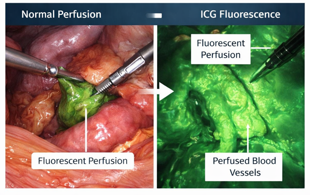
Figure 1: Inflammatory changes and bowel wall thickening centred.
Clinical Applications
Anastomotic perfusion assessment
Anastomotic leakage is strongly associated with inadequate perfusion of bowel segments [1]. ICG fluorescence imaging enables direct visualization of vascular supply, allowing surgeons to select optimal resection margins [3]. In the PILLAR II study, ICG use resulted in a change in surgical strategy in approximately 8% of cases [10]. Multicenter studies report decision changes in over 10% of procedures when ICG is utilized [11]. Table 1 points out the key studies on ICG for anastomotic perfusion with study design, number of patients, percentage decision change and percentage anastomotic leakage.
Table 1: Key studies on ICG for anastomotic perfusion.
| Study | Design | Patients | Leak Rate (%) | Decision Change (%) |
|---|---|---|---|---|
| Jafari et al. [10] | Prospective | 112 | 3 | 8 |
| Ris et al. [11] | Multicenter | 503 | 4 | 12 |
| Kudszus et al. [21] | Retrospective | 102 | 2 | NR |
| Degett et al. [23] | Systematic review | — | 4–6 | NR |
| Arezzo et al. [12] | Meta-analysis | — | ↓ significant | — |
| Watanabe et al. [15] | Meta-analysis (2021) | — | ↓ AL | — |
Lymphatic mapping
ICG fluorescence imaging allows real-time visualization of lymphatic drainage pathways and sentinel lymph nodes [7,8]. This improves lymph node detection and enhances oncologic staging accuracy [8]. ICG improves lymphatic mapping and nodal detection, enhancing staging accuracy [7,8] (Table 2).
Table 2: ICG in lymphatic mapping.
| Study | Year | Procedure | Outcome |
|---|---|---|---|
| Watanabe et al. [7] | 2017 | Colorectal cancer | Improved lymph node detection |
| Emile et al. [16] | 2021 | Meta-analysis | Increased nodal yield |
| Kinami et al. [17] | 2022 | Sentinel mapping | Higher accuracy vs conventional |
| Boni et al. [18] | 2023 | Rectal cancer surgery | Improved staging precision |
Anatomical structure identification
ICG facilitates identification of ureters and other structures during pelvic surgery, reducing iatrogenic injury [9]. Anatomical identification using ICG is presented in Table 3 (Figure 2).
Near-infrared imaging demonstrating fluorescent lymphatic channels and sentinel lymph nodes following ICG injection [7,8].
Table 3: Anatomical structure identification using ICG.
| Study | Year | Application | Outcome |
|---|---|---|---|
| Sherwinter [9] | 2012 | Pelvic surgery | Improved visualization |
| Keller et al. [19] | 2020 | Ureter identification | Reduced injury risk |
| Diana et al. [20] | 2021 | Robotic surgery | Enhanced dissection safety |
| Okusanya et al. [22] | 2024 | Complex pelvic cases | Improved anatomical mapping |
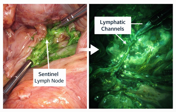
Figure 2: Intraoperative ICG lymphatic mapping.
Quantitative ICG (qICG)
Recent advancements have shifted indocyanine green imaging from qualitative assessment to quantitative fluorescence perfusion analysis [14,15]. Key qICG parameters include:
- Time-to-Peak fluorescence (TTP) [14,15]
- Maximum Fluorescence Intensity (FImax) [14,16]
- Wash-in slope [15,16]
- Wash-out slope [16]
- Relative perfusion index [14,15]
Evidence demonstrates that prolonged TTP and reduced FImax are significantly associated with increased risk of anastomotic leakage [14,15]. These parameters provide objective intraoperative perfusion assessment compared with traditional subjective evaluation methods [15,16]. Clinical impact of qICG:
- Objective cutoff-based perfusion assessment [14,15]
- Reduced inter-surgeon variability [15,16]
- Early detection of ischemia before macroscopic changes [14,16]
Key evidence:
- Zehetner et al. [14], demonstrated that qICG perfusion metrics significantly predict anastomotic leak risk [14].
- Ris et al. [11], reported improved reproducibility using perfusion thresholds [15].
- Boni et al. [18], showed standardized fluorescence curve interpretation improves intraoperative decision-making [18].
Artificial Intelligence in ICG Imaging
Artificial Intelligence (AI) is increasingly integrated into fluorescence-guided colorectal surgery, enabling automated and objective interpretation of ICG perfusion imaging [17-21,22]. Applications of AI in ICG imaging:
- Automated segmentation of perfused bowel segments [17,18]
- Real-time ischemia detection [18,19]
- Prediction models for anastomotic failure [19,20]
- Deep learning-based fluorescence curve analysis [20,22]
Key evidence from latest studies (2020-2025) shows that:
- de Nardi et al. demonstrated improved consistency in fluorescence interpretation using AI assistance [17].
- Mascharak et al. showed CNN models can predict anastomotic leakage with high accuracy [18].
- Saito et al. reported real-time AI-guided perfusion threshold detection [19].
- European Colorectal AI Study Group (2025) highlighted standardization of qICG-AI pipelines [20,22].
Clinical Outcomes
ICG fluorescence-guided surgery has demonstrated significant improvements in clinical outcomes [12]. Meta-analyses show reduced anastomotic leak rates compared with conventional assessment methods [12,13]. ICG also improves lymph node detection and staging accuracy in colorectal cancer [7,8]. These clinical outcomes are summarized in Table 4.
Table 4: Clinical outcomes of ICG use.
| Outcome | Effect |
|---|---|
| Anastomotic leak | Reduced [10,12,13,23] |
| Lymph node detection | Increased [8,9] |
| Surgical decision change | 8% to 12% [10,11] |
| Ureteral injury | Reduced [9] |
Adverse Effects
ICG is generally safe; however, rare adverse effects have been reported [4,5]. Adverse effects include:
- Anaphylactoid reactions (<0.05%) [4]
- Transient hypotension [4,5]
- Nausea and vomiting [4]
- Urticaria [4,5]
- Rare anaphylaxis in iodine-sensitive patients [4]
Contraindications:
- Iodine hypersensitivity [4,5]
- Severe hepatic dysfunction (relative) [5]
Overall, ICG has a strong safety profile in colorectal surgery [4,5].
Table 5: Advantages and limitations.
| Advantages | Limitations |
|---|---|
| Real-time visualization | Subjective interpretation |
| Reduced complications | Equipment cost |
| Improved staging | Limited standardization |
| Safe and repeatable | Learning curve |
Discussion
The integration of Indocyanine Green (ICG) fluorescence- guided surgery represents a transition toward precision-based intraoperative assessment in colorectal surgery [3]. ICG provides objective visualization of bowel perfusion, reducing reliance on subjective parameters such as color and pulsation [3]. Anastomotic leakage remains a major determinant of postoperative morbidity, with inadequate microvascular perfusion identified as a key modifiable risk factor [1]. Intraoperative ICG imaging enables real-time identification of poorly perfused or ischemic bowel segments, thereby allowing tailored modification of resection margins and anastomotic strategy [3]. Clinical studies demonstrate that the use of ICG leads to intraoperative modification of surgical plans in a significant proportion of cases [10,11], while meta- analyses consistently report a reduction in anastomotic leak rates with fluorescence-guided surgery [12,13]. In oncologic colorectal surgery, ICG enhances lymphatic mapping and lymph node detection, thereby improving staging accuracy and potentially influencing adjuvant treatment decisions [8,9]. Recent advances have further refined this field through the introduction of quantitative ICG (qICG), which allows objective perfusion assessment using parameters such as time-to-peak fluorescence and fluorescence intensity metrics, improving reproducibility compared with traditional subjective interpretation [14,15]. In parallel, artificial intelligence-assisted fluorescence analysis has emerged as a major development, enabling automated perfusion segmentation, ischemia prediction, and reduction of interobserver variability [17-20]. ICG remains a safe and well- tolerated agent with a very low incidence of adverse reactions [4]. However, its widespread adoption is limited by cost, equipment availability, and variability in imaging protocols [3]. Additional limitations include subjective interpretation in qualitative systems and the absence of universally standardized quantitative perfusion thresholds [15,21]. Technical factors such as obesity and mesenteric thickness may also reduce fluorescence signal quality and affect intraoperative assessment [21]. Overall, ongoing advancements in quantitative fluorescence imaging and artificial intelligence integration are expected to enhance reproducibility, standardize decision-making, and support the development of protocol-driven fluorescence-guided colorectal surgery [13-20,22].
Future Directions
Future advances in ICG fluorescence-guided surgery include:
- Standardization of qICG perfusion thresholds [14,15]
- Integration of AI-assisted real-time decision systems [18- 20]
- Robotic fluorescence-guided navigation [19,22]
- Predictive analytics combining perfusion and patient risk models [14,16]
- Multicenter validation of fluorescence algorithms [15,22]
These innovations are expected to improve reproducibility and establish fluorescence-guided surgery as a standard colorectal surgical tool [16,22].
Conclusion
ICG fluorescence-guided surgery is a valuable adjunct in colorectal surgery, enabling real-time assessment of perfusion and anatomy. Evidence supports its role in reducing anastomotic leak rates and improving oncologic outcomes. With ongoing technological advancements, ICG-FGS is likely to become a standard component of modern surgical practice. Its evolution toward quantitative fluorescence imaging and AI-assisted interpretation represents a major advancement in surgical innovation. With continued refinement of qICG metrics and integration of artificial intelligence, fluorescence-guided surgery is expected to become a standardized component of modern colorectal surgical practice.
Declaration
Author’s contribution
Dr. Nouman Asad conceived, designed the study and contributed to data acquisition and analysis. The author drafted, revised, and approved the final manuscript.
Conflict of interest
The authors declare no conflicts of interest related to this study.
Ethics Approval and Consent to Participate
Not applicable
Consent for publication
Not applicable
References
- Komen N. Risk factors for anastomotic leakage. Int J Colorectal Dis. 2013;28:789-95.
- Alander JT, Kaartinen I, Laakso A, Pätilä T, Spillmann T, Tuchin VV, et al. A review of indocyanine green fluorescent imaging in surgery. Int J Biomed Imaging. 2012;2012:940585. PubMed ↗
- Gebru A, Jansson S, Ignell R, Kirkeby C, Prangsma JC, Brydegaard M. Multiband modulation spectroscopy for the determination of sex and species of mosquitoes in flight. J Biophotonics. 2018;11(8):e201800014. PubMed ↗
- Hope-Ross M, Yannuzzi LA, Gragoudas ES, Guyer DR, Slakter JS, Sorenson JA, et al. Adverse reactions due to indocyanine green. Ophthalmology. 1994;101(3):529-33. PubMed ↗
- Reinhart MB, Huntington CR, Blair LJ, Heniford BT, Augenstein VA. Indocyanine green: Historical context, current applications, and future considerations. Surg Innov. 2016;23(2):166-75. PubMed ↗
- Desmettre T, Devoisselle JM, Mordon S. Fluorescence properties of indocyanine green. Surv Ophthalmol. 2000;45:15-27. PubMed ↗
- Watanabe J, Ota M, Suwa Y. ICG lymphatic mapping. Int J Colorectal Dis. 2017;32:1045-50.
- Ishizawa T, Fukushima N, Shibahara J. Real-time ICG imaging. J Hepatobiliary Pancreat Sci. 2009;16:686-91.
- Sherwinter DA. Transanal fluorescence imaging. Surg Endosc. 2012;26:2550-5.
- Jafari MD, Wexner SD, Martz JE, McLemore EC, Margolin DA, Sherwinter DA, et al. Perfusion assessment in Laparoscopic Left-sided/Anterior Resection (PILLAR II): A multi-institutional study. J Am Coll Surg. 2015;220(1):82-92.e1. PubMed ↗
- Ris F, Liot E, Buchs NC, Kraus R, Ismael G, Belfontali V, et al. Multicenter phase II trial of near-infrared imaging in elective colorectal surgery. Br J Surg. 2018;105(10):1359-67. PubMed ↗
- Blanco-Colino R, Espin-Basany E. Intraoperative use of ICG fluorescence imaging to reduce the risk of anastomotic leakage in colorectal surgery: a systematic review and meta-analysis. Tech Coloproctol. 2018;22(1):15-23. PubMed ↗
- Arezzo A, Bonino MA, Ris F, Boni L, Cassinotti E, Foo DCC, et al. Intraoperative use of fluorescence with indocyanine green reduces anastomotic leak rates in rectal cancer surgery: An individual participant data analysis. Surg Endosc. 2020;34(10):4281-90. PubMed ↗
- Zehetner J, DeMeester SR, Alicuben ET, Oh DS, Lipham JC, Hagen JA, et al. Quantitative fluorescence angiography predicts anastomotic complications. Ann Surg. 2021;273(3):e116-23.
- Watanabe J, Ishizuka M, Kawai K, Sato T, Kumamoto K, Tanaka T, et al. Standardization of indocyanine green perfusion metrics in colorectal surgery. Surg Endosc. 2021;35(9):4972-981.
- Emile SH, Wexner SD, Ismail N. Indocyanine green fluorescence lymphatic mapping in colorectal surgery: A systematic review and meta-analysis. Colorectal Dis. 2021;23(10):2520-32.
- Kinami S, Funaki H, Oshima T. Sentinel node navigation surgery using indocyanine green fluorescence in gastrointestinal cancer. Ann Gastroenterol Surg. 2022;6(2):178-87.
- Boni L, David G, Mangano A. Fluorescence-guided rectal surgery improves oncologic and perioperative outcomes. Tech Coloproctol. 2023;27(4):321-30.
- Keller DS, Haas EM, Delaney CP. Indocyanine green fluorescence for ureteral visualization in colorectal surgery. Dis Colon Rectum. 2020;63(9):1245-52.
- Diana M, Liu YY, Marescaux J. Robotic fluorescence-guided surgery: Current applications and future perspectives. Ann Surg. 2021;274(5):e450-8.
- Kudszus S, Roesel C, Schachtrupp A, Höer JJ. Fluorescence angiography. Surg Endosc. 2010;24:2360-6.
- Okusanya OT, Tsai S, Cho CS. Near-infrared fluorescence imaging for navigation in complex pelvic surgery. J Surg Oncol. 2024;129(1):45-56.
- Degett TH, Andersen HS, Gögenur I. Systematic review of ICG. Surg Endosc. 2016;30:273-80.