Delayed Diagnosis of Tuberculous Meningitis in an Elderly Male: A Case Report
Abstract
Background
Tuberculous Meningitis (TBM) remains a significant global health concern, with over 164,000 cases occurring annually worldwide with a mortality rate of approximately 48%. Despite England's status as a country with low rates of tuberculosis, recent data shows a concerning 11% increase in TB notifications in 2023, with extrapulmonary forms such as TBM presenting particular di- agnostic challenges. The diagnosis of TBM in elderly patients presents additional challenges due to atypical presentations and reduced inflammatory responses.
Case Report
We report the case of a male in his early 70s who presented with a 14-day history of severe headache, fever, and con- stitutional symptoms following recent travel to Pakistan. His initial presentation mimicked viral infection, which contributed to diag- nostic delays. The patient subsequently developed communicating hydrocephalus and neurological deterioration, ultimately requiring neurosurgical intervention. Cerebrospinal fluid analysis revealed characteristic findings of TBM with 100% lymphocytes, an elevated protein of 6 g/L (0.15 g/L - 0.4 g/L), and low glucose of 1.4 mmol/L (2.5 mmol/L - 4.4 mmol/L), though TB PCR was initially negative.
Conclusion
This case highlights the diagnostic challenges of TBM in elderly patients, particularly following travel to high-burden countries. Early recognition of risk factors, maintaining high clinical suspicion despite negative molecular tests, and prompt empirical treatment are crucial for improved outcomes in suspected TBM cases.
Keywords
Tuberculosis-Meningitis; Safety; Case Report; Difficult Diagnosis
Introduction
Tuberculous meningitis is the most severe form of extrapulmonary tuberculosis, accounting for approximately 1.5% of all tuberculosis cases and carrying a mortality rate of 27% even among treated patients [1]. The burden of TBM remains substantial globally, with recent modelling studies estimating 164,000 incident cases annually in adults worldwide, resulting in approximately 78,200 deaths [1].
England has experienced a concerning reversal in tuberculosis control, with 2023 witnessing an 11% increase in TB notifications to 4,855 cases - the largest yearly increase since current reporting began [2-4]. This trend is especially noticeable among individuals born outside the UK, who account for nearly 80% of active TB cases [4]. The rise in travel-associated tuberculosis, especially among those visiting high-burden countries, has emerged as a significant concern [5,6].
TBM poses unique diagnostic challenges due to its insidious onset, nonspecific clinical presentation, and variable laboratory findings [7,8]. These challenges are amplified in elderly patients, who may present with atypical symptoms and demonstrate blunted inflammatory responses [9]. While the geriatric population is considered high risk for tuberculosis, research on TBM in this group is limited. One study found that patients aged 60 and above were nearly 20% more likely to present with altered mental status compared to younger patients, while those under the age of 60 were more likely to experience symptoms such as headache, vomiting, and fever [9].
The condition's diagnostic complexity often leads to treatment delays, contributing to the high morbidity and mortality rates associated with this disease; the same study found their geriatric patients had a 20% decreased survival rate compared to their younger counterparts. The often-ambiguous clinical presentation of TBM in the elderly population is certainly a significant factor in diagnostic delays, and emphasizes the importance of keeping a low threshold for treatment when considering a diagnosis of TBM.
Case Description
Patient information and initial presentationA male in his early 70s presented to the Emergency Department in early 2024, with a 14-day history of feeling unwell. His chief complaint was severe left-sided headache radiating to the back of his head, rated 10/10 in intensity. He had recently returned from Pakistan six days prior to presentation, where he had attended a wedding celebration.
Associated symptoms included palpitations, intermittent fever with rigors (particularly severe in the morning), reduced oxygen saturation when supine, vomiting, increased urination, and reduced appetite. The patient had been prescribed antibiotics in Pakistan for unclear reasons, which provided no symptomatic relief. His general practitioner had recently seen him in late February, and prescribed nitrofurantoin for a suspected urinary tract infection based on his urine analysis. Past medical history included Benign Prostatic Hyperplasia (BPH), gout, and hypertension with the relevance of BPH potentially mimicking genitourinary tuberculosis and hypertension signaling increased intracranial pressure. He was a non-smoker, did not consume alcohol, and lived independently with his family in the UK. He had no known allergies.
Clinical findingsOn initial examination, the patient was alert and oriented with a Glasgow Coma Scale (GCS) of 15/15. Vital signs revealed tachycardia (HR 101 bpm), mild hypoxia (SpO2 96% on room air), hypertension (BP 170/66 mmHg), and fever (38.7°C). Neurological examination showed no focal deficits, with pupils equal and reactive to light. Cardiovascular examination revealed normal heart sounds without murmurs, and respiratory examination was unremarkable with clear lung fields bilaterally.
Importantly, there were no initial signs of meningism, neck stiffness, or photophobia and the patient remained alert and cooperative throughout the initial assessment – all the above findings were supportive of a viral cause for his symptoms.
Timeline and diagnostic assessmentDays 1-3: Initial assessment focused on fever of unknown origin following recent travel to Pakistan. CT head was performed to rule out intracranial hemorrhage given the sudden onset of severe headache, which was reported as normal by the consultant radiologist. The patient was started on empirical IV co-amoxiclav and supportive care.
Days 4-7: Despite antibiotic treatment, the patient continued to spike temperatures and developed signs of systemic illness. CT thorax, abdomen, and pelvis suggested possible pyelonephritis which led to treatment escalating to IV meropenem, and additional investigations were requested including TB ELISpot, TB PCR and HIV testing.
Days 8-11: Bilateral lymphadenopathy was noted on examination, prompting ultrasound of the neck which revealed isoechoic nodules in the thyroid but no abnormal lymph nodes. The patient remained febrile despite broad-spectrum antibiotics, and inflammatory markers remained normal (CRP and procalcitonin within normal limits); TB PCR was also negative.
Days 15-17: TB ELISpot returned positive, raising suspicion for tuberculosis. The family members noticed the patient developing delirium, and neurological examination revealed reduced ability to lift his left forearm and difficulty with eye movements, though no definitive neck stiffness was observed.
Day 17: Urgent CT head revealed acute communicating hydrocephalus with signs of raised intracranial pressure. Given the combination of positive TB ELISpot, clinical deterioration, and hydrocephalus, tuberculous meningitis was strongly suspected.
Cerebrospinal fluid analysisLumbar puncture was initially unsuccessful and ultimately required sedation; opening pressure of less than 4 cm H2O was found. CSF analysis of the yellow straw-colored fluid revealed: Cell count - 100% lymphocytes; Protein: 6.0 g/L (0.15 g/L - 0.4 g/L); Glucose - 1.4 mmol/L (2.5 mmol/L - 4.4 mmol/L) [10,11].
These findings, particularly the lymphocytic pleocytosis with elevated protein and low glucose, were highly suggestive of TBM despite the negative TB PCR [12,13].
Therapeutic interventionBased on the clinical presentation, CSF findings, and positive TB ELISpot, empirical anti-tuberculosis treatment was initiated, including Rifater (combination of rifampicin 120 mg, isoniazid 50 mg, pyrazinamide 300 mg) - 6 tablets daily; Ethambutol 1200 mg daily; Pyridoxine 20 mg daily; and Dexamethasone 8 mg three times daily.
Additional supportive measures included ceftriaxone and antivirals to cover potential bacterial and viral meningitis, along with aspirin 300 mg daily for its potential neuroprotective effects [14].
Neurological complicationsA brain MRI revealed a left pontine and thalamic ischemic infarct, consistent with TB vasculitis - a well-recognized complication occurring in 15% to 57% of TBM patients [14,15]. The patient developed right-sided upper limb weakness and expressive dysarthria, indicating involvement of the pontine and thalamic regions (Figure 1, 2).
Neurosurgical managementDue to progressive hydrocephalus, the patient was referred to the nearest specialist tertiary center where an Ommaya reservoir with wide-bore external ventricular drain was inserted to manage the communicating hydrocephalus. A sample taken from the shunt confirmed the diagnosis with a positive PCR. This intervention was necessary as hydrocephalus occurs in up to 85% of TBM cases and often requires surgical management when medical therapy fails (Figure 3) [16,17].
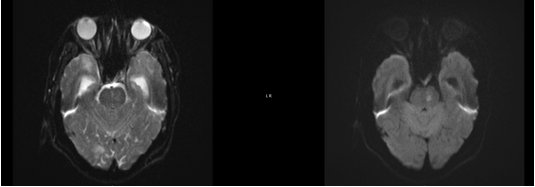
Figure 1: An MRI of the brain showing a left pontine infarct.
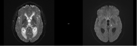
Figure 2: An MRI of the brain showing a left thalamic infarct.
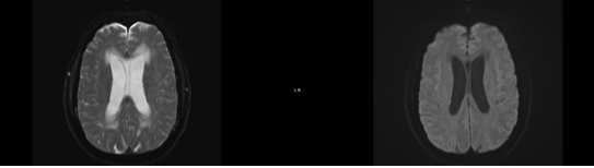
Figure 3: An MRI of the brain showing hydrocephalus.
Follow-up and outcomesThe patient's hospital course was complicated by the following:
- Pulmonary embolism and pneumonia in mid-April, managed with dalteparin and IV tazocin
- Significant weight loss (16% over 3 weeks)
- Oral thrush requiring antifungal treatment
- Deranged liver function tests secondary to anti-TB medications
The patient showed gradual improvement with physiotherapy and occupational therapy. Nasogastric feeding was discontinued on towards the end of April, as oral intake improved. He was discharged in mid-May, with ongoing anti-TB treatment and regular follow-up.
Telephone follow-up in July 2024 confirmed good compliance with medications and no new concerns. The patient was scheduled to complete 12 months of anti-TB treatment, with clinic discharge planned for March 2025.
Discussion
This case exemplifies the diagnostic challenges associated with tuberculous meningitis, particularly in elderly patients following travel to high prevalence countries. Several key learning points emerge from this case, particularly the importance of maintaining high clinical suspicion despite negative molecular tests, and prompt empirical treatment.
Diagnostic challenges in elderly patientsTBM in elderly patients presents unique diagnostic challenges due to altered immune responses and atypical clinical presentations [9]. In this case, the patient's initial presentation with severe headache and fever following travel to Pakistan raised suspicion for various infectious causes, but the absence of classical meningeal signs and negative test results initially delayed the diagnosis. Research indicates that elderly TBM patients more frequently present with altered mental status (96.4% vs. 78.1% in younger patients) while fever, headache, and vomiting are paradoxically less common [9].
The cerebrospinal fluid findings in our patient were classic for TBM, with lymphocytic pleocytosis (100% lymphocytes), markedly elevated protein (6.0 g/L; reference 0.15 g/L - 0.4 g/L), and low glucose (1.4 mmol/L; reference 2.5 mmol/L - 4.4 mmol/L). These findings, particularly protein levels >1 g/L and glucose <2.2 mmol/L, have good specificity for TBM diagnosis, though sensitivity remains limited [10,11,13].
Travel-associated tuberculosis riskThe patient's recent travel to Pakistan, a country with high TB burden (276 cases per 100,000 population annually), represented a significant risk factor [18]. Pakistan has a particularly high burden of multidrug-resistant tuberculosis, making accurate diagnosis and appropriate treatment selection crucial [19]. Travel to high-burden countries increases the risk of acquiring latent TB infection, with greater risk associated with prolonged travel and higher TB burden in the destination country [5,6].
Molecular diagnostic limitationsThe negative TB PCR result in this case highlights the limitations of molecular diagnostics in TBM diagnosis. GeneXpert MTB/RIF, while rapid and specific, has a sensitivity of only approximately 50% for TBM [7]. This underscores the importance of clinical judgment and comprehensive diagnostic approaches when TBM is suspected, particularly in high-risk patients.
Management considerationsThe prompt initiation of empirical anti-TB treatment despite negative molecular tests proved crucial in this case. The combination of four-drug anti-TB therapy (rifampicin, isoniazid, pyrazinamide, ethambutol) with adjunctive corticosteroids represents standard care for TBM. Dexamethasone has been shown to reduce mortality in TBM patients by approximately 25%, though its effect on preventing severe disability remains uncertain [13].
The development of hydrocephalus, occurring in up to 85% of pediatrics TBM cases and commonly in adults, necessitated neurosurgical intervention [16]. The use of external ventricular drainage followed by Ommaya reservoir placement provided effective CSF diversion while allowing for ongoing monitoring and treatment.
Cerebrovascular complicationsThe development of pontine and thalamic infarcts in our patient represents a common but serious complication of TBM. These infarcts typically result from TB vasculitis affecting the basal ganglia, internal capsule, and thalamus - areas commonly referred to as the "tubercular zone" [15]. The presence of cerebral infarction is an independent predictor of mortality in TBM patients, with an adjusted odds ratio of 2.1 for death [20].
Interestingly, conventional cardiovascular risk factors including age, hypertension, and Diabetes mellitus are important predictors of cerebral infarction in TBM patients, suggesting a complex pathophysiology involving both inflammatory and atherothrombotic mechanisms [20].
Clinical implicationsThis case reinforces several important clinical principles:
- High index of suspicion: TBM should be considered in patients with subacute neurological symptoms, especially those with relevant travel history or risk factors
- Empirical treatment: Negative molecular tests should not preclude treatment initiation when clinical suspicion is high
- Multidisciplinary approach: Management of TBM requires coordination between infectious disease specialists, neurologists, and neurosurgeons
- Vigilance for complications: Regular monitoring for hydrocephalus, cerebral infarction, and drug-related adverse effects is essential
Conclusion
This case demonstrates the diagnostic complexities and clinical challenges associated with TBM in elderly patients. The combination of atypical presentation, negative initial molecular tests, and travel-associated risk factors created a diagnostic challenge that required high clinical suspicion and prompt empirical treatment. The diagnostic delay in this case was a result of a multitude of factors, including the vague presentation and symptoms (or lack thereof) that better supported other diagnoses. The patient presented without typical meningeal signs and was initially treated for a suspected UTI; diagnosis was further complicated by an initial lack of elevated inflammatory markers and a negative TB PCR. The decline in the patient’s cognitive function also initially went unnoticed as the staff did not realize he was previously fluent in English, and they did not recognize that his inability to communicate in the language was a change from baseline. The patient's ultimate recovery, despite significant complications, underscores the importance of early recognition and aggressive management of this potentially fatal condition.
Key learning points include the need for maintaining high clinical suspicion in patients with relevant travel history and risk factors, the limitations of molecular diagnostics in TBM diagnosis, and the importance of prompt empirical treatment initiation. The case also highlights the multidisciplinary approach required for optimal TBM management and the need for vigilant monitoring for complications including hydrocephalus and cerebrovascular events.
As global travel continues to increase and tuberculosis rates rise in certain regions, clinicians must remain vigilant for travel- associated tuberculosis and maintain awareness of the diagnostic challenges posed by this serious condition. Early diagnosis and appropriate treatment remain crucial for improving outcomes in this potentially devastating disease.
References
- Dodd PJ, Osman M, Cresswell FV, Stadelman AM, Lan NH, Thuong NTT, et al. The global burden of tuberculous meningitis in adults: A modelling study. PLOS Glob Public Health. 2021;1(12):e0000069. PubMed ↗
- UK Health Security Agency. Executive summary. 2025. Gov.uk ↗
- UK Health Security Agency. With Tuberculosis (TB) on the rise again, how can we prevent further spread? UK Health Security Agency. 2025. UKHSA ↗
- UK Health Security Agency. Tuberculosis incidence and epidemiology, England, 2023. UK Health Security Agency. 2025. Gov.uk ↗
- Hayward S, Harding RM, McShane H, Tanner R. Factors influencing the higher incidence of tuberculosis among migrants and ethnic minorities in the UK. F1000Res. 2018;7:461. PubMed ↗
- Brophy J. Summary of the statement on international travellers who intend to visit friends and relatives. Can Commun Dis Rep. 2015;41(5):89-99. PubMed ↗
- Ghimire B, Thapaliya I, Yadav J, Bhandari S, Paudyal MB, Mehta N, et al. Diagnostic challenges in tuberculous meningitis: A case report with negative GeneXpert result. Ann Med Surg (Lond). 2023;85(11):5731-5. PubMed ↗
- Foppiano Palacios C, Saleeb PG. Challenges in the diagnosis of tuberculous meningitis. J Clin Tuberc Other Mycobact Dis. 2020;20:100164. PubMed ↗
- Rahman N, Pannu AK, Yadav R, Sethi S, Saroch A, Garg M, et al. Tuberculous meningitis in the elderly. QJM. 2022;115(6):381-7. PubMed ↗
- Hrishi AP, Sethuraman M. Cerebrospinal Fluid (CSF) analysis and interpretation in neurocritical care for acute neurological conditions. Indian J Crit Care Med. 2019;23(Suppl 2):S115-S9. PubMed ↗
- Leen WG, Willemsen MA, Wevers RA, Verbeek MM. Cerebrospinal fluid glucose and lactate: Age-specific reference values and implications for clinical practice. PLoS One. 2012;7(8):e42745. PubMed ↗
- Nand L, Chauhan PS, Sharma S, Shandil R. Spectrum of cerebrospinal fluid analysis and nervous system diseases in hospitalised patients: A hospital based prospective study from a hilly state of Himachal Pradesh in North India. Int J Adv Med. 2020;7(4):571-6. PubMed ↗
- Solomons RS, Visser DH, Donald PR, Marais BJ, Schoeman JF, van Furth AM. The diagnostic value of cerebrospinal fluid chemistry results in childhood tuberculous meningitis. Childs Nerv Syst. 2015;31(8):1335-40. PubMed ↗
- Misra UK, Kalita J, Maurya PK. Stroke in tuberculous meningitis. J Neurol Sci. 2011;303(1-2):22-30. PubMed ↗
- Tai MLS, Viswanathan S, Rahmat K, Nor HM, Kadir KAA, Goh KJ, et al. Cerebral infarction pattern in tuberculous meningitis. Sci Rep. 2016;6(1):38802. PubMed ↗
- Paliwal VK, Garg RK. Hydrocephalus in tuberculous meningitis – pearls and nuances. Neurol India. 2021;69(Suppl 2):S330-5. PubMed ↗
- Rajshekhar V. Management of hydrocephalus in patients with tuberculous meningitis. Neurol India. 2009;57(4):368. PubMed ↗
- Jawed A, Tharwani ZH, Siddiqui A, Masood W, Qamar K, Islam Z, et al. Better understanding extrapulmonary tuberculosis: A scoping review of public health impact in Pakistan, Afghanistan, India, and Bangladesh. Health Sci Rep. 2023;6(6):e1357. PubMed ↗
- Pakistan: Migrant health guide. Public Health England. GOV.UK. 2017. Gov.uk ↗
- Wasay M, Khan M, Farooq S, Khowaja ZA, Bawa ZA, Mansoor Ali S, et al. Frequency and impact of cerebral infarctions in patients with tuberculous meningitis. Stroke. 2018;49(10):2288-93. PubMed ↗