In Vivo Efficacy of Ethanol Extract from Peliosanthes Micrantha Rhizomes on Serum Testosterone and Sexual Behavior Parameters in Male Sprague-Dawley Rats
Abstract
Peliosanthes micrantha species is traditionally employed in the Dak Nong province of Vietnam for its supposed properties in improving male sexuality. The objective of this study was to evaluate the impact of P. micrantha ethanolic rhizome extract administration on sexual performance in inexperienced male sprague-dawley rats. The following sexual performances were evaluated: body weights, weights of reproductive organs, and levels of testosterone for repeated doses of 7 days; several parameters including mount behaviors such as Mount Frequency (MF), Mount Latency (ML); intromission behaviors such as Intromission Frequency (IF), Intromission Latency (IL), Intromission Efficiency (IE); ejaculation behaviors such as Ejaculation Frequency (EF), Post Ejaculatory Interval (PEI), Ejaculation Latency (EL); and Copulatory Efficiency behavior (CE) for single dose and repeated doses of 7 days. Results from the 7 days repeated-dose experiment showed increases in body weights, up to 9.00%, slight changes in reproductive organs' weights, and a significant increase in serum testosterone levels in extract-treated rats. The tested extracts showed different effects of sexual behavior parameters between acute single doses and subacute repeated doses during 7 days of P. micrantha rhizomes ethanolic extract administration. Our results indicated that the repeated and high doses have better effects in Mount Latency (ML), and Intromission Frequency (IF). On the contrary, the repeated and low doses have better effects in Intromission Latency (IL).
Keywords
Peliosanthes micrantha; Rhizome; Ethanolic extract; Testosterone; Sexual behavior; Male sprague-dawley rats
Introduction
Erectile Dysfunction (ED) is a common disorder affecting the quality of life of millions of male patients and their partners. Nowadays, ED is increasingly viewed as a biopsychosocial disorder with an intricate etiology and a range of diverse available therapeutic approaches. It is estimated that over 150 million men worldwide experience ED to varying degrees [1]. Numerous studies have demonstrated that ED is closely associated with various comorbidities and risk factors, including aging, reduced androgen levels, obesity, and cardiovascular disease [2,3]. Some current therapies to treat ED include oral Phosphodiesterase 5 Inhibitor (PDE5I) drugs, intracavernosal and intraurethral administration of erectogenic injections, hormonal replacement therapy, etc. [4]. Sexual dysfunction refers to the inability to achieve or maintain normal sexual function, encompassing partial or complete erectile failure, difficulty in sustaining an erection, premature ejaculation, decreased libido, orgasmic disorders, arousal disturbances, and impaired tumescence. Previous studies have demonstrated that penile erection largely depends on the relaxation of penile arteries and erectile tissues, particularly the smooth muscle of the corpus cavernosum [5]
As the primary male sex hormones, androgens are essential for the differentiation and sustained expression of the male phenotype during embryonic development, puberty, and adult life. As the major androgen in circulation (~90%), testosterone is synthesized in Leydig cells of the testes and metabolized by 5α-reductase to generate the more biologically active 5α-dihydrotestosterone. Disturbances of testosterone impair spermatogenesis, male secondary sexual characteristics development, sexual performance, and subsequent male infertility. The abnormally low serum testosterone levels are generally characterized by male erectile dysfunction. Restoring serum testosterone to normal physiological levels may improve or resolve these clinical manifestations [6,7]. Appropriate testosterone concentrations are crucial for proper endothelial function, as the expression of penile PDE5 isoenzyme. Current drugs available, including sildenafil, tadalafil, vardenafil, and avanafil, which are used to treat ED, can cause side effects [8-10]. It has been shown that traditional medicines can enhance sexual activity without side effects [11]. A study using the male Wistar rats showed that the administration of Allium tuberosum seed extract improved corpus cavernosum smooth muscle relaxation and sexual behavior parameters [12]. A recent study on the female rats for 21 consecutive days using up to 400 mg/kg of A. americana extract showed the significant enhancement of sexual behaviors, including mounting and intromission frequencies, libido activity, erection rate, and both quick and long flip responses [13]. 70% ethanolic extract of Garcinia kola on male sexual function model treated daily for 56 days orally with 100 mg/kgP, 200 mg/kgP, and 400 mg/kgP showed increased testicular weights, increased serum testosterone levels [14]. Many studies have also shown androgens, with the most well-known testosterone compound, as male sexual hormones presented by medicinal plants [15]. The Tribulus terrestris extract showed androgen-enhancing aphrodisiac and pro-erectile effects from experimental animal studies [16]. The acetone extracts of Peltophorum africanum and the methanol extract of Terminalia sambesiaca have had good androgenic properties as their testosterone productions were at 0.147 and 0.188 ng/mL, respectively [17].
The P. micrantha Aver et N. Tanaka, sp. nov. (called Sam cau Krông Nô or Hue da la nho in Vietnamese) was found in central highland Vietnam [18]. According to the indigenous people in Krong No district, Dak Nong province, Vietnam, the rhizomes of P. micrantha were traditionally alcohol extracted and have been used to increase physical endurance, and for male impotence. Only a few studies of chemistry and biological activities have been published for the P. micrantha species. In the alcoholic extract of its rhizomes, UHPLC-Q-TOF-MS identified three known compounds, namely pumilaside A, pumilaside C, β–sitosterol, and glycoside J-3. The P. micrantha extract was standardized and showed mild DPPH antioxidant activity [19,20]. However, the effects of P. micrantha on male sexual behavior have not yet been experimentally characterized. Given the increasing clinical interest in plant-derived therapies for managing erectile dysfunction and androgen deficiency, it is essential to establish preclinical evidence before human application. Therefore, the present study is designed to evaluate the in vivo efficacy of the ethanolic extract of P. micrantha rhizomes using a sprague– dawley rat model. Specifically, we investigate both the immediate and 7-day repeated oral administration to determine their impact on both sexual behavior parameters and serum testosterone levels. This approach aims to clarify whether the observed aphrodisiac potential is primarily mediated through endocrine modulation, behavioral enhancement, or a combination of both mechanisms, thereby providing a translational basis for future clinical investigations.
Materials and Methods
Chemicals and reagentsEstradiol benzoate, thiopental, and testosterone were purchased from Sigma-Aldrich Chemical (St. Louis, MO, USA). Progesterone was procured from Glentham Life Sciences (Germany). EliKine Testosterone ELISA kit was from Abbkine Co. (Michigan, USA). Analytical-grade ethanol and other reagents were procured from Merck (USA).
Plant material and extract preparationThe rhizomes of P. micrantha were collected in Dak Nong Province, Vietnam, in October 2021 and identified by Dr. KHANG Nguyen Sinh of the Institute of Ecology and Biological Resources (IEBR), VAST. The voucher specimen (PM-R-2021) has been lodged at the CHTD, VAST. After washing fresh rhizomes of P. micrantha (8.70 kg), the materials were sliced and dried in a hot-air oven at 80°C for 24 h, and finally milled into fine powder.
Dried powder of the rhizomes of P. micrantha (2.80 kg) was extracted thrice with 96% ethanol (17.5 L) at room temperature under ultrasonication in a 20 kHz and 1 kW extractor as described previously [19]. The combined filtrates were vacuum- concentrated to obtain a brown solid residue, referred to as PM- R-E (123,30 g). The PM-R-E extract was purged with nitrogen and kept at −20°C until further use.
Experimental animalsSixty 12 to 13-week-old Sprague-Dawley rats (60 males and 50 females; 200-250g) were obtained from the National Institute of Hygiene and Epidemiology, Vietnam. Housing conditions and experimentation were in the standard laboratory conditions at 25°C ± 2°C, relative humidity of 44% to 56%, and 12:12 hours of light and dark cycles. They maintained 5 days before the experiment in the Viptam Institute of Technology Application (Viptam). Each rat was individually identified by marking a specific number on its tail with a permanent marker. The design and performance of animal experiments were approved by the Viptam's Ethical Committee (reference number No. 210814/Vip/ Pre).
Sexual behavior studyFemale rats were anesthetized by thiopental and xylazine hydrochloride, ovariectomized, and kept fed during 14 days. Then, they were brought into oestrous by sequential subcutaneous injection with estradiol benzoate (0.25 mg/kg body weight) and followed with progesterone (2.50 mg/kg body weight) at 52 h and 4 h before aphrodisiac studies, respectively [12]. Sexual receptivity was assessed by pairing them with sexually experienced adult males that were not used in the main experiment. The female rat was brought out and replaced by another one if she did not accept the male, and only those showing copulatory behavior were used in the study. Before the experiment, males exhibiting reduced sexual performance were identified through preliminary screening. Briefly, the male rats were placed singly with sexually receptive females for 4 min to 5 min before the exposures, then trained by exposing them to sexually receptive females once daily in the dark cycle for 3 consecutive times with an interval of 4 days. In the present study, we used the male rats that failed to ejaculate in any of the last three mating sessions and were classified as sexually inactive [21].
In our unpublished data, the preliminary hard capsule combination was developed, containing 500 mg of total extract, including 230 mg of P. micrantha ethanolic extract, with intended daily doses of 3 or 6 capsules. The Animal Equivalent Dose (AED, mg/kg) was calculated from the Human Equivalent Dose (HED, mg/kg) using body surface area normalization, according to the following equation:
AED (mg/kg) = HED (mg/kg) × Km (1),
where the Km conversion factor for rats is 6.2.
Minor adjustments were made to account for differences in average human and rat body weights [22]. Based on this approach, the calculated AEDs were 86 mg/kg and 172 mg/kg body weight, corresponding to the human daily intake of 3 and 6 capsules, respectively.
The group size (n = 11 per group) is selected based on previous studies evaluating sexual behavior parameters in rat models, where comparable sample sizes have been shown to detect treatment-related differences [12]. This sample size was also determined considering practical feasibility and ethical principles for animal use, aiming to provide sufficient statistical power for behavioral and hormonal assessments. A total of 44 sexually inexperienced and sluggish male rats were selected, housed separately, and randomly divided into four groups. Lot 1, Normal Control (NC) (n=11): were given Na-CMC 0.5%, 10 ml/kgP. Lot 2, positive control (n=11): were subcutaneously injected with testosterone, 0.4 mg/kgP, 2 times per week. Lot 3 (n=11): was given with the extract at a dose of 280 mg/kgP in Na-CMC 0.5%. Lot 4 (n=11): was given with the extract at a dose of 562 mg/ kgP in Na-CMC 0.5%. Oral administration of the extracts or drugs was performed once daily for 7 days via a metal oropharyngeal cannula. Following a 10 min acclimation period, a sexually receptive female was placed into the male's cage for behavioral assessment. Body weights were recorded before the experiment and again on day 7, while food and water intake were monitored daily throughout the study.
Measurement of sexual parametersSexual performance parameters were evaluated for 30 min by an observer blinded to treatment conditions, during the reversed dark phase, using a dim red light for illumination. An 8MP camera recorded all the activities. The following sexual behavior parameters were analyzed, including Mount Frequency (MF) as the total number of mounts preceding ejaculation at a specified period, Mount Latency (ML) as the time interval between the placement of the female into the cage and the male's first mount accompanied by pelvic thrusting, Intromission Frequency (IF) as the total number of intromissions occurring before ejaculation, Intromission Latency (IL) as the time (in seconds) elapsed from the introduction of the female into the cage to the first mount accompanied by vaginal penetration (intromission), Ejaculation Frequency (EF) as the total number of ejaculations recorded from the time the female was introduced to the male within the specified observation period, Post-Ejaculatory Interval (PEI) as the time elapsed between ejaculation and the subsequent intromission, and Ejaculation Latency (EL) as the time interval (in seconds) between the first intromission and ejaculation. Tests were terminated immediately after the first post-ejaculatory intromission, or if intromission did not occur within 15 min of female introduction, or if ejaculation latency exceeded 30 min.
Two rating behavior indexes were computed as follows:
Copulatory efficiency = (number of intromissions/number of mounts) × 100 (%) or CE= (IF/MF) × 100 (%);
Intromission efficiency = number of intromissions/(number of mounts + number of intromissions) × 100 (%) or
IE = IF/(IF+MF) × 100 (%) [23].
Measurement of testosterone levels and reproductive organs' weightsAt the end of the treatment period, all rats were weighed and sacrificed. Following cardiac puncture, serum samples were isolated and preserved at −20°C prior to quantification of testosterone levels by ELISA in accordance with the manufacturer's guidelines. Reproductive organs, including testis, seminal vesicles, bulbourethral glands, levator ani, prostate glands, and glans penis, were collected immediately after dissection by open castration. Thereafter, samples were dissected free of surrounding tissues and fat, rinsed thrice with physiological saline, and blotted dry using filter paper to remove remaining blood and fluid. The weights of dried organs were determined.
Data analysisData were presented as mean ± Standard Deviation (SD), along with the minimum and maximum values. All statistical analyses were performed with SPSS version 16.0 (SPSS Inc., Chicago, IL, USA), two-way Analysis of Variance (ANOVA), and further analyzed using Dunnett's multiple comparison test and Fisher's LSD test. Frequency comparison tables of the parameters of sexual behaviors were generated along with the p-value from Fisher's exact test (Chi–square) and assessed for statistical significance by the Mann-Whitney U-test. Differences were considered statistically significant at p<0.05.
Results
Effects on body weights, reproductive organs' weights, and testosterone levelsAttention was given to body weight evaluation before and at the end of the experiment, as presented in Table 1. Figure 1 shows the comparative body weights of the rats between the 0th and 7th days of the experiment, taking Lot 1 as the base. After 7 days of the repeated-dose experiments, the mean weights of the dosed rats were not statistically different in comparison to NC. Results showed that all the increases in body weights were observed for all the groups of tested animals till the end of treatment (day 7). In body weights, the control rats (NC), the positive control rats (P), and two extract-dosed rats (PM-R-E-1 and PM-R-E-2) showed significant increases (~2.27%, 5.50%, 9.00%, and 8.40%, respectively (p<0.05).
Table 1: Weighting evaluation of body weights in tested rats.
| Lot/dose (mg/kg) | Body weight (g) | Body weight change (%) | |
|---|---|---|---|
| Before administration | 7th day | ||
| Lot 1, normal control (NC): Na-CMC 0.5% | 244.44 ± 13.02 | 250.00 ± 11.67 | 2.27 |
| Lot 2, positive control: testosterone, 0.4 mg/kgP | 234.28 ± 14.77 | 247.14 ± 20.55 | 5.49 |
| Lot 3: PM-R-E-1, 86 mg/kgP | 230.10 ± 10.75 | 250.67 ± 1.03 | 8.94 |
| Lot 4: PM-R-E-2, 172 mg/kgP | 227.00 ± 10.40 | 246.00 ± 13.21 | 8.37 |
*p<0.05 means that the difference is statistically significant
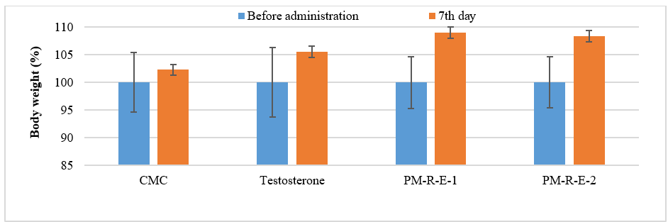
Figure 1: Dose-dependent body weight change in tested rats.
After 7 days of the repeated-dose experiments, mean weight changes in the reproductive organs of the tested rats in different experimental groups were presented in Table 2. Figure 2 shows the comparative weights of different reproductive organs in the tested rats, taking Lot 1 as the base. Results showed that the weights of the seminal vesicle, levator ani, prostate glands, and glans penis from all the rats significantly increased in comparison to NC rats (p <0.05). Both of the PM-R-E–treated animal groups presented higher weight increase than the P rats. Adversely, the testicle weights of the PM-R-E-1 and testosterone–treated animal groups were increased, but one of the PM-R-E-2–treated rats was decreased in comparison to NC rats. The weights of bulbourethral glands in both of the PM-R-E–treated animal groups were not changed as compared to NC rats.
Table 2: Weighting evaluation of reproductive organs in tested rats.
| Lot/dose (mg/kg) | Weight of organs (g/100gP rats) | |||||
|---|---|---|---|---|---|---|
| Testicles | Seminal vesicle | Bulbourethral glands | Levator ani | Prostate glands | Glans penis | |
| Lot 1, NC: Na-CMC 0.5% | 0.74 ± 0.07 | 0.45 ± 0.14 | 0.03 ± 0.01 | 0.34 ± 0.06 | 0.10 ± 0.04 | 0.05 ± 0.01 |
| Lot 2, P: testosterone, 0.4 mg/kgP | 0.81 ± 0.12 | 0.48 ± 0.07 | 0.02 ± 0.01 | 0.38 ± 0.06 | 0.10 ± 0.02 | 0.07 ± 0.01 |
| p>0.05 | p>0.05 | p>0.05 | p>0.05 | p>0.05 | p<0.01 | |
| Lot 3: PM-R-E-1, 86 mg/kgP | 0.82 ± 0.06 | 0.51 ± 0.1 | 0.03 ± 0.01 | 0.4 ± 0.04 | 0.12 ± 0.03 | 0.06 ± 0.01 |
| p<0.01 | p>0.05 | p>0.05 | p<0.05 | p>0.05 | p>0.05 | |
| Lot 4: PM-R-E-2, 172 mg/kgP | 0.71 ± 0.09 | 0.48 ± 0.09 | 0.03 ± 0.01 | 0.41 ± 0.08 | 0.13 ± 0.03 | 0.06 ± 0.01 |
| p>0.05 | p>0.05 | p<0.05 | p<0.05 | p>0.05 | p>0.05 | |
*p<0.05 means that the difference is statistically significant
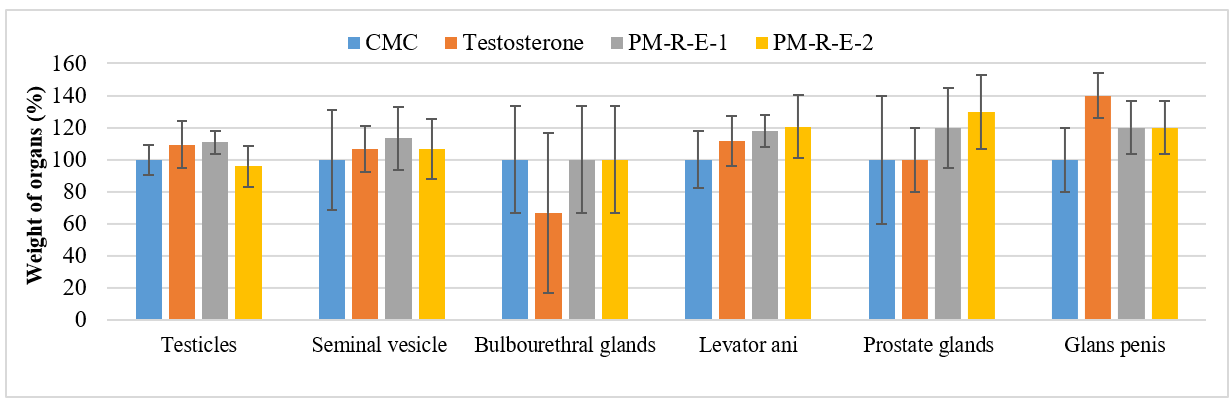
Figure 2: Comparative different organ weights in each experimental group.
The serum testosterone concentrations of the treated rats were quantified by ELISA, and the findings are shown in Table 3 and Figure 3. After 7 days of the repeated-dose experiments, the mean testosterone levels increased significantly with the testosterone- and PM-R-E-2–treated animal groups at 172 mg/kgP, while the lower dose of PM-R-E-1 at 86 mg/kgP decreased serum testosterone concentrations.
Table 3: Serum testosterone levels in tested rats.
| Lot/dose (mg/kg) | Testosteron (ng/dl) | |
|---|---|---|
| Before administration | 7 days | |
| Lot 1, normal control (NC): Na-CMC 0.5% | 15.12 ± 3.54 | 6.92 ± 1.78 (p>0.05) |
| Lot 2, positive control (P): testosterone, 0.4 mg/kgP | 16.16 ± 3.31 | 49.23 ± 1.87 (p<0.05) |
| Lot 3: PM-R-E-1, 86 mg/kgP | 7.52 ± 1.89 | 2.8 ± 0.71 (p>0.05) |
| Lot 4: PM-R-E-2, 172 mg/kgP | 6.83 ± 1.15 | 12.03 ± 6.23 (p>0.05) |
*p<0.05 means that the difference is statistically significant
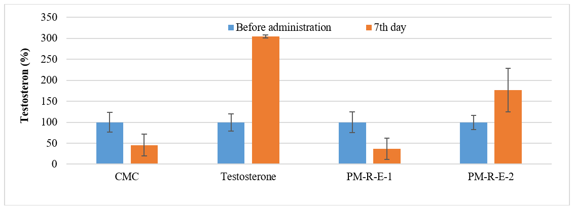
Figure 3: Serum testosterone levels in rats from experimental groups.
Acute single-dose administration effects on sexual behavior parametersAcute sexual behaviors of the tested rats were observed after 2 hours of the extract administration. Behavioral observations revealed proceptivity in female rats, characterized by ear-wiggling, lordosis, and hopping, alongside enhanced sexual activity in treated males. The treated males demonstrated quick orientation toward the females and exhibited characteristic precopulatory behaviors such as body sniffing, anogenital inspection, and circling, ultimately leading to mounting. The results of acute rating behaviors were presented in Table 4 and Figure 4, and showed that the intromissions were observed in all the rats. The mounts were lower in all the treated rats in comparison to the NC rats. However, the ejaculations were slightly higher in the extract- treated rats in comparison to the NC rats.
Table 4: Acute rating behaviours in tested rats.
| Lot/dose (mg/kg): 2 h | Mount (%) | Intromission (%) | Ejaculation (%) |
|---|---|---|---|
| Lot 1, normal control (NC): Na-CMC 0.5% | 72.73 | 100 | 81.82 |
| Lot 2, positive control (P): testosterone, 0.4 mg/kgP | 63.64 | 100 | 63.64 |
| Lot 3: PM-R-E-1, 86 mg/kgP | 27.27 | 100 | 90.91 |
| Lot 4: PM-R-E-2, 172 mg/kgP | 70 | 100 | 90 |
*p<0.05 means that the difference is statistically significant
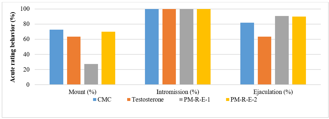
Figure 4: Acute rating behaviours in rats from experimental groups.
In Table 5 and Figure 5, the results obtained from the acute sexual behavior analysis (after 2h) relating to male rat sexual characteristics of both PM-R-E-treated rats indicated that PM- R-E significantly decreased mean ML, MF, and EF when compared with the rats in the untreated NC group (p>0.05). However, the P rats remarkably decreased ML and EF when compared with the rats in the PM-R-E-1 treated group. In addition, the extract also led to a remarkably decreased ML in the rats in the PM-R-E-2 treated group compared to all other tested groups. The results showed a marked increase in the mean IL, IF, and EL values, but a decrease in the PEI values in both of P and PM-R-E-2 treated groups in comparison to the NC rats. Conversely, the extract at the PM-R-E-1 dose decreased the mean IL and IF in comparison to the NC rats. Compared to the NC group, rats receiving treatment showed increased CE and IE indices.
Table 5: Acute sexual behaviours in tested rats.
| Lot/dose (mg/kg) | ML (s) | MF (times) | IL (s) | IF (times/30 min) | EL (s) | PEI (s) | EF (times/30 min) | CE (%) | IE (%) |
|---|---|---|---|---|---|---|---|---|---|
| Lot 1, NC: Na-CMC 0.5% | 132.11 ± 361.02 | 3.56 ± 3.54 | 35.33 ± 46.47 | 45.67 ± 16.39 | 566.11 ± 330.2 | 390.78 ± 56.15 | 2 ± 0.71 | 1282.87 | 92.77 |
| Lot 2, P: testosterone | 74.09 ± 194.55 | 1.73 ± 1.68 | 37.82 ± 37.84 | 58.09 ± 17.82 | 1035.82 ± 708.81 | 330 ± 51.73* | 1.09 ± 0.94* | 3357.8 | 97.11 |
| p=0.65 | p=0.15 | p=0.90 | p=0.13 | p=0.08 | p=0.05 | p=0.03 | |||
| Lot 3: PM-R-E-1, 86 mg/kgP | 108.5 ± 359.34 | 1.09 ± 2.47 | 19.55 ± 21.12 | 41.82 ± 14.46 | 587 ± 456.00 | 415.2 ± 67.19 | 1.73 ± 0.9 | 3836.7 | 97.46 |
| p=0.89 | p=0.08 | p=0.33 | p=0.58 | p=0.91 | p=0.40 | p=0.47 | |||
| Lot 4: PM-R-E-2, 172 mg/kgP | 49.1 ± 5.82 | 2.27 ± 2.53 | 67.27 ± 94.48 | 60.00 ± 17.63 | 600.19 ± 495.36 | 383.3 ± 218.66 | 1.91 ± 1.14 | 2643.17 | 96.35 |
| p=0.26 | p=0.36 | p=0.37 | p=0.08 | p=0.83 | p=0.92 | p=0.84 |
*p<0.05 means that the difference is statistically significant
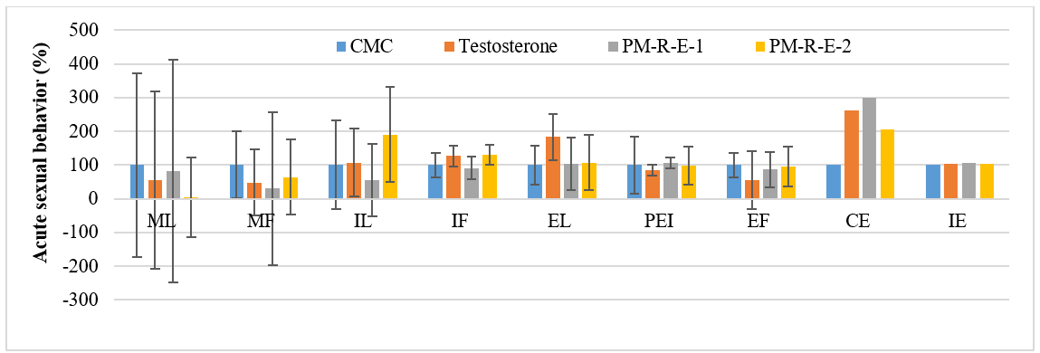
Figure 5: Acute sexual behaviours in rats from experimental groups.
Subacute multi-dose administration effects on sexual behavior parametersSubacute sexual behaviors of the tested rats were observed after 7 days of repeated extract administration. The results of subacute rating behaviors were presented in Table 6 and Figure 6, and showed that the intromissions were observed in all the tested rats. The mounts were lower in all the extract-treated rats in comparison to the NC rats. The ejaculations were remarkably lower in the lower extract dose-treated rats in comparison to the NC rats. However, the ejaculations were observed in all the higher extract dose-treated rats.
Table 6: Subacute rating behaviours in tested rats.
| Lot/dose (mg/kg): 7 days | Mount (%) | Intromission (%) | Ejaculation (%) |
|---|---|---|---|
| Lot 1, normal control (NC): Na-CMC 0.5% | 100 | 100 | 81.82 |
| Lot 3: PM-R-E-1, 86 mg/kgP | 81.82 | 100 | 54.55 |
| Lot 4: PM-R-E-2, 172 mg/kgP | 90 | 100 | 100 |
*p<0.05 means that the difference is statistically significant
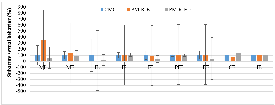
Figure 6: Subacute sexual behaviours in rats from experimental groups.
Table 7 and Figure 6 present the subacute sexual behaviors in tested rats after repeated doses during 7 days. The high mean ML and mean MF values were recorded in the lower extract dose-treated rats administered at 86 mg/kgP while the lower values were observed in the higher extract dose-treated rats in comparison to NC rats. The treatment duration with the extract had no significant effect on either ML or MF, as revealed by the two-way ANOVA analysis (p>0.05). There is a remarkable decrease in the mean IL and a somewhat higher increase in the mean IF of the rats administered with the extracts at 86 mg/ kgP and 172 mg/kgP in comparison to NC rats. According to the statistical analysis, no significant differences in mean IL and IF were observed across treatment durations (p>0.05). It was observed that the mean EL was remarkably increased in the rats administered with the extract at 172 mg/kgP, and the mean EF was slightly changed at the dose of 86 mg/kgP in comparison to NC rats. As the statistical ANOVA parameter p<0.05, the higher extract dose had a significant effect on both EL and EF during the treatment duration. The mean PEI values were shown to be higher at the rats administered with both doses of extract at 86 mg/kgP and 172 mg/kgP in comparison to NC rats.
Table 7: Subacute sexual behaviours in tested rats.
| Lot/dose (mg/kg) | ML (s) | MF (times) | IL (s) | IF | EL (s) | PEI (s) | EF (times/30 min) | CE (%) | IE (%) |
|---|---|---|---|---|---|---|---|---|---|
| Lot 1, NC: Na-CMC | 75.00 ± 120.33 | 4.82 ± 3.03 | 94.82 ± 255.54 | 42.00 ± 20.21 | 872.91 ± 600.55 | 390.13 ± 84.12 | 1.45 ± 0.93 | 871.37 | 89.71 |
| Lot 3: PM-R-E-1, 86 mg/kgP | 264.22 ± 498.11 | 6.44 ± 5.39 | 11.56 ± 6.11 | 43.44 ± 22.57 | 830.22 ± 740.41 | 439.5 ± 56.80 | 1.56 ± 1.24 | 674.53 | 87.09 |
| p=0.24 | p=0.4 | p=0.34 | p=0.88 | p=0.89 | p=0.24 | p=0.84 | |||
| Lot 4: PM-R-E-2, 172 mg/kgP | 41.20 ± 73.54 | 3.9 ± 3.57 | 20.8 ± 19.78 | 44.80 ± 13.17 | 359.50 ± 213.10* | 391.9 ± 82.16 | 2.20 ± 0.63* | 1148.72 | 91.99 |
*p<0.05 means that the difference is statistically significant
Discussion
The growing demand for herbal medicines as aphrodisiacs stems from the perception that they are safe, natural, and associated with minimal adverse effects. The ethnobotany knowledge alluded to the use of numerous plants/natural products/combinations, including P. micrantha, as sex enhancers in the absence of robust scientific research. To substantiate these traditional claims scientifically, the ethanolic extract of P. micrantha rhizomes was studied for the pharmacological effects on the serum testosterone and sexual behavior parameters using the male Sprague-Dawley rat model in this study. Our data analysis indicated marked increases in body weights, up to 9.00% over 7 days of the experiment in all the tested animal groups. Body weight changes were considered an indicator of physiological and metabolic reactions within an organism. Weight gain is often associated with an increase in both anabolic and metabolic processes, while in most cases, weight loss is believed to be associated with disease conditions. Changes in body weight can be influenced by, but not limited to, feed consumption and disease conditions. Thus, the body weight changes in the experiments may lead to poor male reproductive health [24]. The increased weights of reproductive organs were observed in a previous in vivo aphrodisiac activity study of the Aquilaria malaccensis extract, suggesting its spermatogenic potential [25].
Our serum testosterone analysis showed a divergent effect of P. micrantha extract, with the lower dose at 86 mg/kg of PM- R-E-1 dose resulting in a reduction, while the higher dose at 172 mg/kg of PM-R-E-2 dose produced an increase. This apparent inconsistency may be attributed to several factors. First, inter- individual variability among animals, particularly in hormonally sensitive endpoints such as testosterone, may have contributed to fluctuations within groups, especially given the relatively small sample size [26]. Second, the response may reflect a non-linear or biphasic dose–response relationship, which is commonly observed in phytochemical-based interventions, where low doses may exert inhibitory or negligible effects, while higher doses activate stimulatory pathways [27]. From a mechanistic perspective, it is plausible that at higher concentrations, the extract enhances Leydig cell steroidogenesis, potentially via modulation of intracellular signaling pathways (e.g., cAMP- mediated processes), leading to increased testosterone synthesis [28]. In contrast, lower doses may be insufficient to activate these pathways or may transiently influence regulatory feedback mechanisms within the hypothalamic–pituitary–gonadal axis, resulting in reduced circulating testosterone levels [29]. The reduced testosterone level was observed in Leydig cells treated with the extracts of Camellia sinensis and Aspalathus linearis.
Spermatogenesis is mainly controlled by the male reproductive system, and its fertilization is ensured by the delivery of spermatozoa to the female reproductive tract. In our study, the reproductive organ weights were assessed. The testicular weights of the P rats and PM-R-E-1 extract-treated rats were increased as compared with those of NC rats, while the values of the PM-R-E-2 extract-treated rats were decreased. For the Aquilaria malaccensis extract, the changes in testicular weight may reflect alterations in the seminiferous tubules, where a decrease or increase corresponds to reduced or enhanced spermatogenic activity, respectively [25]. The weight changes of other reproductive organs also indicate the changes in physiological and pathological status, as they could influence the function of these organs. The standardized Serenoa repens extract also affected the weight changes of reproductive organs and the spermatogenesis [30]. Thus, the oral administration of P. micrantha extract for 7 days slightly affects reproductive organs' weights.
The androgen-dependent mechanisms may be the cause of the weight changes of reproductive organs. Androgen levels are related to the contents of sexual hormones, including testosterone, in animals. Circulating testosterone plays a crucial role in modulating sexual desire or stimulation. Through the modulation of neurotransmitter systems, especially dopaminergic pathways, these hormones contribute to increased locomotion and improved copulatory efficiency. The circulation of testosterone during copulatory encounters plays a pivotal role in augmenting sexual desire, drive, and functional performance and enhances sexual desire, motivation, and overall sexual function [31]. A significant increase in serum testosterone levels was observed in the PM-R-E-treated group compared to controls. A key role of testicular Leydig cells is the biosynthesis of testosterone. Data from this study imply that PM-R-E exerts testosterone-like effects and may promote Leydig cell steroidogenesis by influencing intracellular cAMP levels in vitro [32]. It was shown that Tribulus cistoides extract strongly increased hormone levels, improved nitric oxide levels, and gonadotropin concentrations by the bioactive phytochemicals' mediation [33].
Acute single-dose studySexual activity was evaluated on day 1 and day 7 following the initiation of extract administration. In our experiments, carried out on young sexually inexperienced rats, we have examined a complete pattern of male rat sexual behavior. Sexual behaviour parameters such as MF, ML, IF, IL, EF, EL, and PEI were recorded after 2 h of the extract administration at the 1st day.
Latency parameters such as Mount Latency (ML), Ejaculation Latency (EL), Intromission Latency (IL), and Post-Ejaculatory Interval (PEI) serve as reliable parameters for evaluating sexual motivation in male rats. A reduction in ML and IL reflected a shortened interval between the introduction of a receptive female and the onset of mounting or intromission behavior. The observed decline in latency implies heightened sexual responsiveness and provides evidence for the Mirabilis jalapa extract's potential aphrodisiac action [34]. In the present study, PM-R-E at both tested doses (86 mg/kg and 172 mg/kg) and testosterone injection significantly decreased ML and PM-R-E-1 of the 86 mg/kg dose only significantly decreased IL as compared with the NC rats. The PM-R-E-2 extract-treated group also led to a remarkably decreased ML compared to all other tested groups. Consequently, the observed decline in ML and IL supports the efficacy of the extract in promoting sexual motivation and behavioral performance in male rats. Our results corroborate previous findings obtained with Massularia acuminata root treatment at doses of 25 mg/kg to 100 mg/kg in male Wistar rats [35]. Conversely, an increase in another latency parameter, Ejaculation Latency (EL), indicates higher sexual motivation. The EL parameter reflects a significant enhancement in copulatory performance in both male and female rats [36,37]. Results of the present study showed an increase in EL values of P and PM- R-E-2 treated rats in comparison to the NC rats. The least EL values were shown by the rats in the PM-R-E-1 treated group. The prolongation of the EL by PM-R-E-2 treated rats suggests an aphrodisiac action. A decrease in Post-Ejaculatory Interval (PEI) in male rats indicates an increase in potency and libido [38]. Our study showed the decrease in the PEI of both P and PM- R-E-2 treated groups in comparison to the NC rats, suggesting enhanced sexual vigor and a higher engagement in subsequent sexual activity.
The Ejaculation Frequency (EF), Intromission Frequency (IF), and Mount Frequency (MF) are useful sexual frequency indices of vigor, libido, and potency. There was a significant increase in the number of MF that reflects sexual motivation. Moreover, the efficiency of erection, penile orientation, and easy ejaculatory reflexes are shown by an increase in the IF frequency. As intromission is not possible without adequate erection and coordinated activity of penile muscles, the increase in IF suggests that the mechanism of penile erection was activated. Since an increase in EF is an indication of enhanced aphrodisiac effect, the presence of plug in the vagina of the female rats indicates that ejaculation occurs [39]. In our study, PM-R-E significantly decreased MF and EF when compared with the rats in the untreated NC group (p>0.05). However, the P rats remarkably decreased EF when compared with the rats in the PM-R-E-1 treated group. In comparison to the NC rats, there was a marked increase in the IF values at both of the P and PM-R-E-2 treated groups, and, conversely, the extract at the PM-R-E-1 dose decreased the IF values. The increase in IF following the PM-R-E-2 administration suggests improved sexual libido. Previous study using the Syzygium aromaticum's ethanolic extract in male rats showed the same results [40].
The mounting and intromission behaviors without any inhibition of Mount Frequency (MF), Intromission Frequency (IF), Copulatory Efficiency (CE), or Intromission Ratio (IE) were exhibited by all the rats. These findings showed that libido, sexual vigor, and overall sexual performance remained unimpaired during the aphrodisiac effect of the extract. In our results, the intromission percentages were the same (100%) in all the rats administered with the extracts and testosterone. Ejaculation percentages were slightly increased in all the rats administered the extracts. The lowest mount percentages were recorded in rats administered the PM-R-E-1 dose. Both the CE and IE indices were higher in treated rats relative to the NC rats. The prolonged EL of the PM-R-E extract at both doses is an indication that CE performance in the rats was enhanced. Similar performances have been observed on the extracts of Allium tuberosum [41] and Moringa oleifera [42]. All the present data showed that acute aphrodisiac administration of P. micrantha rhizome ethanolic extract improved the performance of sexually inexperienced male rats.
Subacute multi-dose studyThe sexual activity test was observed using one repeated dose per day till the 7th day. In our experiments, the ML and MF were significantly increased by PM-R-E-1 at 86 mg/kgP ML and decreased by PM-R-E-2 at 172 mg/kgP dose as compared with the NC rats. The decrease in ML following repeated dosing indicates the efficacy of the extract in enhancing sexual motivation and performance in male rats. The efficacy of the extract to enhance sexual performance and motivation of male rats was shown by the decrease in ML following repeated dosing. This finding is consistent with the results reported for the aqueous extract of the M. angustifolia aerial parts administered at a dose of 300 mg/ kg for 7 days to male Wistar rats [43]. The remarkable decreases in the mean IL were observed for the rats administered with both doses of the extracts. The repeated-dose study showed a somewhat higher increase in mean IF of the rats administered with the extracts at 86 mg/kgP and 172 mg/kgP in comparison to NC rats. The IF increase suggests improved sexual vigor. Similar results were found while working on Boesenbergia rotunda in male rats [44]. The observation of the remarkably increased mean EL values in the rats administered with the extract at 172 mg/kgP indicates direct sexual intercourse between male and female rats, bypassing mounting and intromission and leading to ejaculation as found for Rauvolfia vomitoria ethanolic extract on sexual performance in male rats [45]. Our results of long administration duration till 7 days showed the mean PEI was higher in the extract-treated rats, contradicting the fact that the decrease in PEI in male rats presents an increase in a faster recovery rate from exhaustion during sexual intercourse [38]. As the mean EF values were observed to be slightly higher at the dose of 86 mg/kgP, this indicated the enhanced aphrodisiac effect. Similar performances have been observed on the ethanol extracts of Salvia haematodes on reproductive function and copulatory behavior in male rats [46].
Our behavioral study indicated that the effects of acute single doses and subacute repeated doses during 7 days of P. micrantha rhizomes ethanolic extract administration were different. For comparisons between the high dose group and the normal control group, the study showed the same decreasing trends in Mount Latency (ML), Mount Frequency (MF); the same increasing trend in Intromission Frequency (IF); and the slight change in Post Ejaculatory Interval (PEI); but adverse trends in Intromission Latency (IL), Ejaculation Latency (EL), and Ejaculation Frequency (EF). Meanwhile, for comparisons between the low dose group and the normal control group, the results showed the same decreasing trend in Intromission Latency (IL); the same increasing trend in Post Ejaculatory Interval (PEI); but adverse trends in Mount Latency (ML), Mount Frequency (MF), Intromission Frequency (IF), Ejaculation Latency (EL), and Ejaculation Frequency (EF). The results meant that the repeated doses at high dose have better effects in Mount Latency (ML), and Intromission Frequency (IF) than at low dose, but worse effects in Mount Frequency (MF). On the contrary, the repeated doses at low dose have better effects in Intromission Latency (IL) than at low dose, but worse effects Post Ejaculatory Interval (PEI).
In a previous study, key phytochemicals, including β-sitosterol and pumilaside A were identified in the ethanolic extract of P. micrantha rhizomes [47]. These compounds have been associated with aphrodisiac and androgenic activities through multiple potential mechanisms. The β–sitosterol isolated from the hexane extract of Mondia whitei may contribute to enhanced steroidogenesis by serving as a precursor or modulator within the testosterone biosynthetic pathway. In addition, β-sitosterol has been reported to improve erectile function in sexually naïve rats, possibly through modulation of Nitric Oxide (NO)-mediated vasodilation and improved penile blood flow [48]. Pumilaside A, identified in Ficus pumila fruits, has been implicated in the co- management of hypertension and erectile dysfunction, suggesting a role in vascular regulation, potentially via endothelial function and smooth muscle relaxation [49,50]. Furthermore, other classes of phytochemicals commonly present in medicinal plant extracts, such as saponins, alkaloids, and flavonoids, may act synergistically to enhance sexual function. These compounds are known to promote vasodilation, increase nitric oxide bioavailability, and modulate hormonal balance, thereby supporting both erectile physiology and androgenic activity [33]. Collectively, these findings suggest that the observed effects of P. micrantha extract may be mediated through a combination of endocrine modulation and peripheral vascular mechanisms, rather than a single pathway, which may also explain the dose- dependent and variable responses observed in the present study.
Conclusion
The present study demonstrates that the ethanolic extract of Peliosanthes micrantha rhizomes enhanced potential aphrodisiac activity in male Sprague–Dawley rats, as evidenced by improvements in selected sexual behavior parameters and modulation of serum testosterone levels following acute and subacute administration. These findings suggest that the extract may exert potent aphrodisiac properties through a combination of endocrine regulation and peripheral mechanisms associated with sexual function. However, several limitations should be acknowledged. The relatively small sample size (n=11 per group) may limit the statistical power and contribute to inter-individual variability in behavioral and hormonal outcomes. In addition, the short treatment duration (7 days) may not fully capture the long-term pharmacological effects or safety profile of the extract. Furthermore, the study did not include histological examination of reproductive organs or comprehensive biochemical analyses (e.g., oxidative stress markers, nitric oxide levels, or key enzymes involved in steroidogenesis), which restricts mechanistic interpretation.
Future studies are therefore warranted to validate and extend these findings. Specifically, investigations with larger sample sizes and longer treatment durations should be conducted to confirm efficacy and reproducibility. Detailed mechanistic studies, including evaluation of hypothalamic–pituitary–gonadal axis regulation, NO signaling pathways, and oxidative stress parameters, are needed to elucidate the underlying modes of action. In addition, histological assessment of testicular and accessory reproductive tissues would provide important insights into structural and functional changes associated with treatment. Such studies will be essential to support the translational potential of P. micrantha as a candidate for managing male sexual dysfunction.
Author Contribution
L.N. Hung: Conceptualization, design of experiments, and draft manuscript. D.N. Thuy: Conceptualization, design, collection and treatment of herbal materials. N.T. Huong: Collection and pre- treatment of herbal materials. L.M. Ha and P.V. Trung: Herbal extracts. Do Thi Nguyet Que: Aphrodisiac experiments. Mai Van Nam: Final manuscript.
Acknowledgment
The project was supported by VAST [Grant Number: UDNDP.04/2022-2023].
Animal Rights Statement
The Ethical Committee, Viptam Institute of Technology Application approved the protocols by reference number No. 210814/Vip/Pre.
Declaration of AI Technology Usage
The authors declare that no Artificial Intelligence (AI) technologies or AI-assisted tools were utilized in any capacity during the writing and preparation of this article.
References
- Wang CM, Wu BR, Xiang P, Xiao J, Hu XC. Management of male erectile dysfunction: From the past to the future. Front Endocrinol (Lausanne). 2023;14:1148834. PubMed ↗
- Zeleke M, Hailu D, Daka D. Erectile dysfunction and associated factors among diabetic patients at Hawassa, southern Ethiopia. BMC Endocr Disord. 2021;21(1):139. PubMed ↗
- Fujita N, Okamoto T, Yamamoto H, Yoneyama T, Hashimoto Y, Mikami T, et al. Association between sex hormones and erectile dysfunction in men without hypoandrogenism. Sci Rep. 2024;14(1):13433. PubMed ↗
- Raheem OA, Natale C, Dick B, Reddy AG, Yousif A, Khera M, et al. Novel treatments of erectile dysfunction: Review of the current literature. Sex Med Rev. 2021;9(1):123-32. PubMed ↗
- Sangiorgi G, Cereda A, Benedetto D, Bonanni M, Chiricolo G, Cota L, et al. Anatomy, pathophysiology, molecular mechanisms, and clinical management of erectile dysfunction in patients affected by coronary artery disease: A review. Biomedicines. 2021;9(4):432. PubMed ↗
- Heemers HV, Tindall DJ. Androgen Receptor (AR) coregulators: A diversity of functions converging on and regulating the AR transcriptional complex. Endocr Rev. 2007;28(7):778-808. PubMed ↗
- Van den Broeck T, Soebadi MA, Falter A, Raets L, Duponselle J, Lootsma J, et al. Testosterone induces relaxation of human corpus cavernosum tissue of patients with erectile dysfunction. Sex Med. 2020;8(1):114-9. PubMed ↗
- Morelli A, Filippi S, Mancina R, Luconi M, Vignozzi L, Marini M, et al. Androgens regulate phosphodiesterase type 5 expression and functional activity in corpora cavernosa. Endocrinology. 2004;145(5):2253-63. PubMed ↗
- Aversa A, Duca Y, Condorelli RA, Calogero AE, La Vignera S. Androgen deficiency and phosphodiesterase type 5 expression changes in aging male: Therapeutic implications. Front Endocrinol (Lausanne). 2019;10:225. PubMed ↗
- Pyrgidis N, Mykoniatis I, Haidich AB, Tirta M, Talimtzi P, Kalyvianakis D, et al. Effect of phosphodiesterase-type 5 inhibitors on erectile function: An overview of systematic reviews and meta-analyses. BMJ Open. 2021;11(8):e047396. PubMed ↗
- Wada AS, Jatau AI, Shitu Z, Hassan MA, Alshargi O, Isa AM, et al. The use of traditional medicines for sexual enhancement in Northern Nigeria. J Community Health. 2023;48(4):670-7. PubMed ↗
- Tang X, Olatunji OJ, Zhou Y, Hou X. In vitro and in vivo aphrodisiac properties of the seed extract from Allium tuberosum on corpus cavernosum smooth muscle relaxation and sexual behavior parameters in male Wistar rats. BMC Complement Altern Med. 2017;17(1):510. PubMed ↗
- Kumar S, Tandel B, Paul A. Evaluation of aphrodisiac activity of Agave americana leaves in male Wistar rats. Int J Pharm Sci Drug Res. 2023;15(1):19-25. IJPSdronline ↗
- Sewani-Rusike CR, Ralebona N, Nkeh-Chungag BN. Dose- and time-dependent effects of Garcinia kola seed extract on sexual behaviour and reproductive parameters in male Wistar rats. Andrologia. 2016;48(3):300-7. PubMed ↗
- Janeczko A. Estrogens and androgens in plants: The last 20 years of studies. Plants (Basel). 2021;10(12):2783. PubMed ↗
- Neychev V, Mitev V. Pro-sexual and androgen enhancing effects of Tribulus terrestris L.: Fact or fiction. J Ethnopharmacol. 2016;179:345-55. PubMed ↗
- Masuku NP, Unuofin JO, Lebelo SL. Phytochemical content, antioxidant activities and androgenic properties of four South African medicinal plants. J Herbmed Pharmacol. 2020;9(3):245-56. PubMed ↗
- Averyanov LV, Tanaka N. New species of Peliosanthes (Asparagaceae) from Vietnam. Turczaninowia. 2013;16:5-7. Researchgate ↗
- Hung LN, Thuy DN, Thanh TV, Huong NT. Standardization of Peliosanthes micrantha extract and male wellness hard capsule. J Vietnam Rural Dev Sci. 2024;83:44-50.
- Thuy DN, Huong NT, Trung PV, Hung LN, Chuong NDT. Study of Peliosanthes micrantha medicinal plant: UHPLC-Q-TOF-MS analysis of phytochemicals and antioxidant activity. J Food Sci. 2024;6(1):14-25. CARIjournals ↗
- Hull EM, Dominguez JM. Sexual behavior in male rodents. Horm Behav. 2007;52(1):45-55. PubMed ↗
- Nair AB, Jacob S. A simple practice guide for dose conversion between animals and human. J Basic Clin Pharm. 2016;7(2):27-31. PubMed ↗
- Kenmogne H, Koloko B, Hambe C, Domkam J, Ngaha Njila M, Bend E, et al. Effects of aqueous root extract of Carpolobia alba G. Don on sexual behaviour in adult male rats. Andrologia. 2016;48(8):908-14. PubMed ↗
- Sharma AM, Pischon T, Hardt S, Kunz I, Luft FC. Hypothesis: β-adrenergic receptor blockers and weight gain: A systematic analysis. Hypertension. 2001;37(2):250-4. AHAjournals ↗
- Zaidi N, Haron MN, Komilus CF, Lananan F, Chew HH, Yaakub N, et al. Effect of Karas (Aquilaria malaccensis) on male reproductive organs and sperm quality in adult Sprague Dawley rats. Trop Life Sci Res. 2023;34(1):241-59. PubMed ↗
- Kempenaers B, Peters A, Foerster K. Sources of individual variation in plasma testosterone levels. Philos Trans R Soc Lond B Biol Sci. 2008;363(1497):1711-23. PubMed ↗
- Jodynis-Liebert J, Kujawska M. Biphasic dose-response induced by phytochemicals: Experimental evidence. J Clin Med. 2020;9(3):718. PubMed ↗
- Monageng E, Offor U, Takalani NB, Mohlala K, Opuwari CS. A review on the impact of oxidative stress and medicinal plants on Leydig cells. Antioxidants (Basel). 2023;12(8):1559. PubMed ↗
- Wang Y, Chen F, Ye L, Zirkin B, Chen H. Steroidogenesis in Leydig cells: Effects of aging and environmental factors. Reproduction. 2017;154(4):R111-22. PubMed ↗
- Yun JM, Lee M, Kim D, Prasad KS, Eun S, Kim OK, et al. Standardized saw palmetto extract directly and indirectly affects testosterone biosynthesis and spermatogenesis. J Med Food. 2021;24(6):617-25. PubMed ↗
- Besong EE, Akhigbe TM, Ashonibare PJ, Oladipo AA, Obimma JN, Hamed MA, et al. Zinc improves sexual performance and erectile function by preventing penile oxidative injury and upregulating circulating testosterone in lead-exposed rats. Redox Rep. 2023;28(1):2225675. PubMed ↗
- Zhang ZB, Yang QT. The testosterone mimetic properties of icariin. Asian J Androl. 2006;8(5):601-5. PubMed ↗
- Nsah KB, Mogho NMT, Fonkwo NS, Antoine KS. The aphrodisiac and androgenic effects of aqueous and ethanol extracts of Tribulus cistoides (Zygophyllaceae) on nicotine-induced sexual dysfunction in male Wistar rats. Scientific World Journal. 2025;2025:7410030. PubMed ↗
- Rahman AU, Alam F, Rehman ZU, Khan M, Ahmad T. Effects of Mirabilis jalapa L. root extract and sildenafil on paroxetine-induced sexual dysfunction in male rats and characterization of its phytoconstituents by UPLC-MS. S Afr J Bot. 2023;152:240-6. ScienceDirect ↗
- Yakubu MT, Awotunde OS, Ajiboye TO, Oladiji AT, Akanji MA. Pro-sexual effects of aqueous extracts of Massularia acuminata root in male Wistar rats. Andrologia. 2011;43(5):334-40. PubMed ↗
- Cizmeci S, Ongun S, Sarac A, Sel E, Tozburun S, Durmus N. Low frequency neuromuscular electrical stimulation applied to the bulbospongiosus muscle prolongs the ejaculation latency in a rat model. Int J Impot Res. 2023;35(8):701-4. PubMed ↗
- Sotelo-Tapia C, Medina AC, Cortes PM, Hernández-Arteaga E, Hidalgo-Aguirre RM, Guevara MA, et al. Ejaculation latency determines susceptibility to stress in the male rat. Behav Process. 2023;205:104819. PubMed ↗
- Olvera-Roldán EO, Cristóbal-Luna JM, García-Martínez Y, Mojica-Villegas MA, Pérez-Pastén-Borja R, Gutiérrez-Salmeán G, et al. Effects of Spirulina maxima on a model of sexual dysfunction in streptozotocin-induced diabetic male rats. Plants (Basel). 2023;12(4):722. PubMed ↗
- Agmo A. Male rat sexual behavior. Brain Res Protoc. 1997;1(2):203-9. PubMed ↗
- Tajuddin A, Ahmad S, Latif A, Qasmi IA. Effect of 50% ethanolic extract of Syzygium aromaticum (L.) Merr. & Perry. (clove) on sexual behaviour of normal male rats. BMC Complement Altern Med. 2004;4:17. PubMed ↗
- Guohua H, Yanhua L, Rengang M, Dongzhi W, Zhengzhi M, Hua Z. Aphrodisiac properties of Allium tuberosum seeds extract. J Ethnopharmacol. 2009;122(3):579-82. PubMed ↗
- Zade VS, Dabhadkar DK, Thakare VG, Pare SR. Effect of aqueous extract of Moringa oleifera seed on sexual activity of male albino rats. Int J Bio Forum. 2013;5(1):129-40. ResearchTrend ↗
- Fouche G, Afolayan AJ, Wintola OA, Khorombi TE, Senabe J. Effect of the aqueous extract of the aerial parts of Monsonia angustifolia E. Mey. Ex A. Rich., on the sexual behaviour of male Wistar rats. BMC Complement Altern Med. 2015;15:343. PubMed ↗
- Ongwisespaiboon O, Jiraungkoorskul W. Fingerroot, Boesenbergia rotunda and its aphrodisiac activity. Pharmacogn Rev. 2017;11(21):27-30. PubMed ↗
- Koloko BL, Bushra I, Wankeu-Nya M, Ngaha Njila MI, Kenmogne H, Nyonseu Nzeubang DC, et al. In vivo effects of Rauvolfia vomitoria (Apocynaceae) ethanolic extract on sexual performance and reproductive activity in male rats. Andrologia. 2020;52(11):e13857. PubMed ↗
- Bansode FW, Rajendran SM, Singh RK. Dose-dependent effects of ethanol extract of Salvia haematodes Wall roots on reproductive function and copulatory behaviour in male rats. Andrologia. 2015;47(3):266-75. PubMed ↗
- Hung LN, Thuy DN, Huong NT, Nhung PT, Trung PV, Que D. Chemico-physical characterization and cytotoxicity of the Peliosanthes micrantha rhizomes collected in Highland Province Dak Nong of Vietnam. Nat Prod Commun. 2026;21(2):1-12. SAGE ↗
- Watcho P, Zelefack F, Ngouela SA, Nguelefack TB, Kamtchouing P, Tsamo E, et al. Enhancement of erectile function of sexually naive rats by β-sitosterol and α-β-amyrin acetate isolated from the hexane extract of Mondia whitei. Asian Pac J Trop Biomed. 2012;2(4):314-9. ScienceDirect ↗
- Kitajima J, Kimizuka K, Tanaka Y. Three new sesquiterpenoid glucosides of Ficus pumila fruit. Chem Pharm Bull (Tokyo). 2000;48(1):77-80. PubMed ↗
- Ajeigbe OF, Oboh G, Ademosun AO. Ficus plants in the co-management of hypertension and erectile dysfunction. Phytomedicine Plus. 2021;1(4):100096. ScienceDirect↗